5X8N image
Deposition Date 2017-03-03
Release Date 2017-04-19
Last Version Date 2023-11-22
Entry Detail
PDB ID:
5X8N
Title:
Crystal structure of mouse importin-alpha1 bound to the nuclear localization signal of Epstein-Barr virus EBNA-LP protein
Biological Source:
Source Organism:
Host Organism:
Method Details:
Experimental Method:
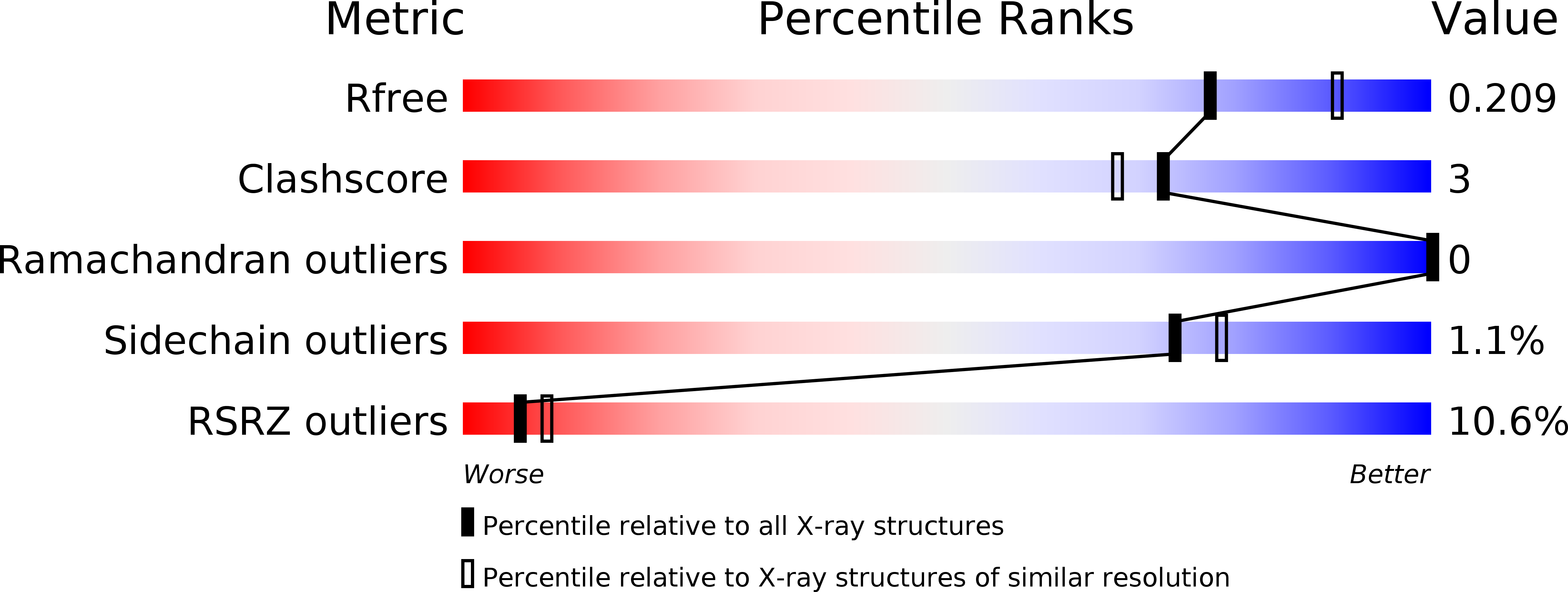
Resolution:
2.15 Å
R-Value Free:
0.20
R-Value Work:
0.18
R-Value Observed:
0.18
Space Group:
P 21 21 21
Macromolecular Entities
Polymer Type:polypeptide(L)
Molecule:Importin subunit alpha-1
Gene (Uniprot):Kpna2
Chain IDs:A
Chain Length:460
Number of Molecules:1
Biological Source:Mus musculus
Polymer Type:polypeptide(L)
Molecule:Epstein-Barr nuclear antigen leader protein
Gene (Uniprot):EBNA-LP
Chain IDs:B
Chain Length:22
Number of Molecules:1
Biological Source:Human herpesvirus 4 (strain B95-8)
Primary Citation
Crystal structure of importin-alpha bound to the nuclear localization signal of Epstein-Barr virus EBNA-LP protein
Protein Sci. 26 1231 1235 (2017)
PMID: 28383161 DOI: 10.1002/pro.3173

Abstact

Epstein-Barr virus EBNA-LP protein is a transcriptional coactivator of EBNA2. Efficient nuclear localization of EBNA-LP is essential for cooperation with EBNA2. Here, we report the crystal structure of the nuclear import adaptor importin-α1 bound to the nuclear localization signal (NLS) of EBNA-LP that shows EBNA-LP residues 44-RRVRRR-49 binding to the major NLS-binding site at the P0-P5 positions. In contrast to previously characterized classical NLSs that invariably have a basic residue [either lysine (in the vast majority of cases) or arginine] at the P2 position, the EBNA-LP NLS is unique in that it has valine at the P2 position. The loss of the critical P2 lysine (or arginine) is compensated by arginine at the P0 position in the EBNA-LP NLS.

Legend

Protein

Chemical

Disease

Primary Citation of related structures