5X7R image
Deposition Date 2017-02-27
Release Date 2017-07-26
Last Version Date 2023-11-22
Entry Detail
PDB ID:
5X7R
Title:
Crystal structure of Paenibacillus sp. 598K alpha-1,6-glucosyltransferase complexed with isomaltohexaose
Biological Source:
Source Organism:
Method Details:
Experimental Method:
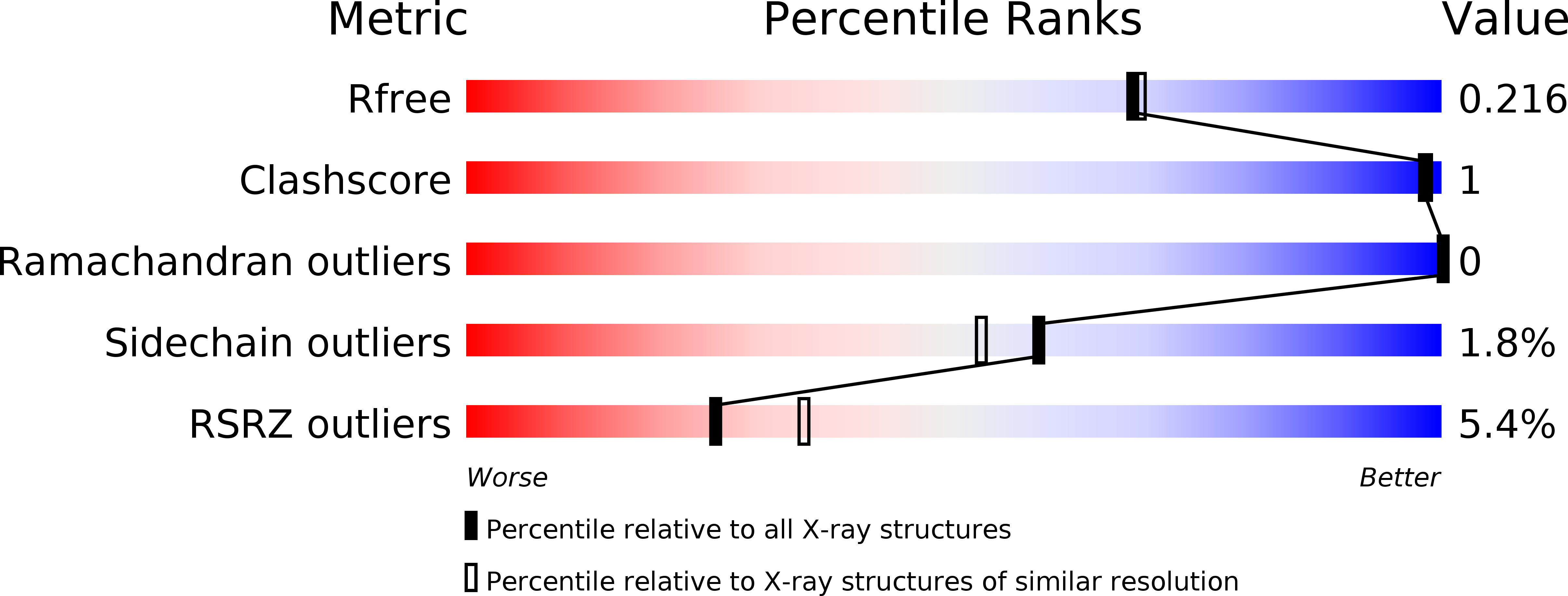
Resolution:
1.95 Å
R-Value Free:
0.20
R-Value Work:
0.17
R-Value Observed:
0.17
Space Group:
C 2 2 21
Macromolecular Entities
Polymer Type:polypeptide(L)
Molecule:Glycoside hydrolase family 31 alpha-glucosidase
Gene (Uniprot):6gt31a
Chain IDs:A, B
Chain Length:1263
Number of Molecules:2
Biological Source:Paenibacillus sp. 598K
Peptide-like Molecules
PRD_900001
PRD_900110
Primary Citation
Carbohydrate-binding architecture of the multi-modular alpha-1,6-glucosyltransferase from Paenibacillus sp. 598K, which produces alpha-1,6-glucosyl-alpha-glucosaccharides from starch
Biochem. J. 474 2763 2778 (2017)
PMID: 28698247 DOI: 10.1042/BCJ20170152

Abstact

Paenibacillus sp. 598K α-1,6-glucosyltransferase (Ps6TG31A), a member of glycoside hydrolase family 31, catalyzes exo-α-glucohydrolysis and transglucosylation and produces α-1,6-glucosyl-α-glucosaccharides from α-glucan via its disproportionation activity. The crystal structure of Ps6TG31A was determined by an anomalous dispersion method using a terbium derivative. The monomeric Ps6TG31A consisted of one catalytic (β/α)8-barrel domain and six small domains, one on the N-terminal and five on the C-terminal side. The structures of the enzyme complexed with maltohexaose, isomaltohexaose, and acarbose demonstrated that the ligands were observed in the catalytic cleft and the sugar-binding sites of four β-domains. The catalytic site was structured by a glucose-binding pocket and an aglycon-binding cleft built by two sidewalls. The bound acarbose was located with its non-reducing end pseudosugar docked in the pocket, and the other moieties along one sidewall serving three subsites for the α-1,4-glucan. The bound isomaltooligosaccharide was found on the opposite sidewall, which provided the space for the acceptor molecule to be positioned for attack of the catalytic intermediate covalent complex during transglucosylation. The N-terminal domain recognized the α-1,4-glucan in a surface-binding mode. Two of the five C-terminal domains belong to the carbohydrate-binding modules family 35 and one to family 61. The sugar complex structures indicated that the first family 35 module preferred α-1,6-glucan, whereas the second family 35 module and family 61 module preferred α-1,4-glucan. Ps6TG31A appears to have enhanced transglucosylation activity facilitated by its carbohydrate-binding modules and substrate-binding cleft that positions the substrate and acceptor sugar for the transglucosylation.

Legend

Protein

Chemical

Disease

Primary Citation of related structures