5X6N image
Deposition Date 2017-02-22
Release Date 2017-03-08
Last Version Date 2024-11-20
Entry Detail
PDB ID:
5X6N
Keywords:
Title:
Structure of P. Knowlesi DBL Domain Capable of binding Human Duffy Antigen
Biological Source:
Source Organism:
Host Organism:
Method Details:
Experimental Method:
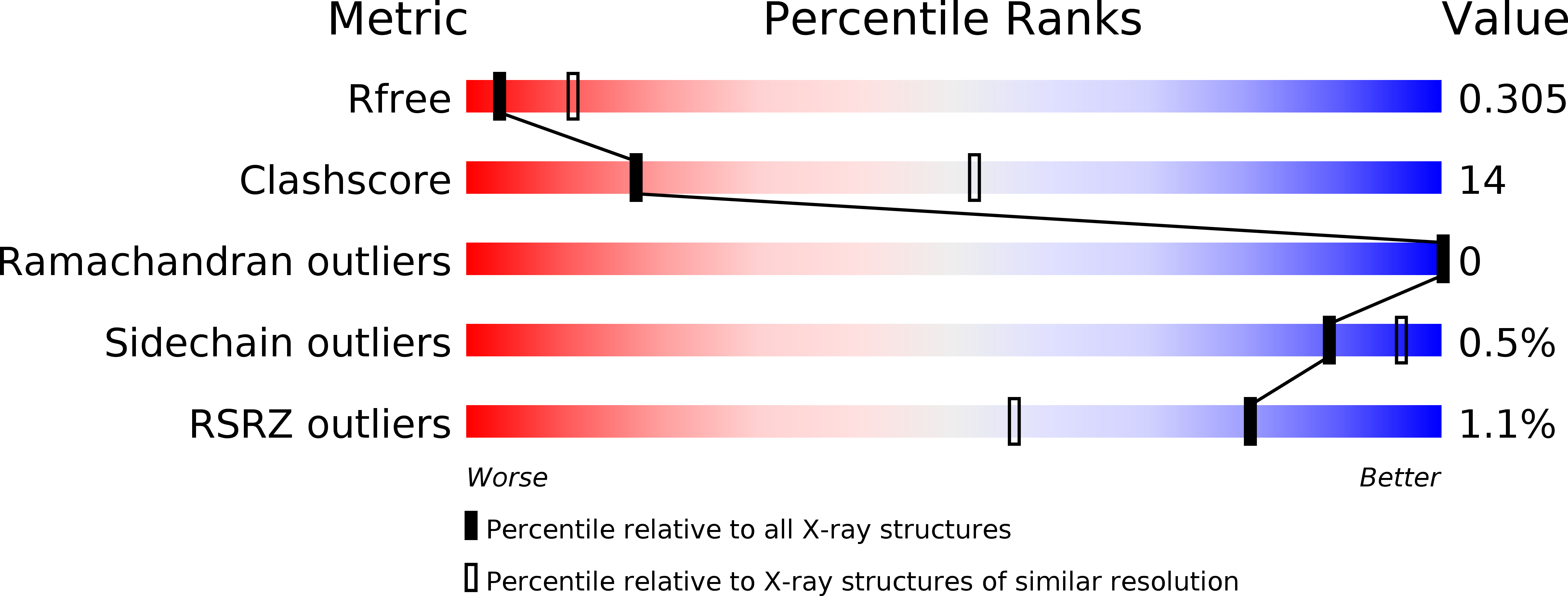
Resolution:
3.00 Å
R-Value Free:
0.29
R-Value Work:
0.24
R-Value Observed:
0.24
Space Group:
P 43 21 2
Macromolecular Entities
Structures with similar UniProt ID
Protein Blast
Polymer Type:polypeptide(L)
Molecule:Duffy binding protein
Chain IDs:A
Chain Length:338
Number of Molecules:1
Biological Source:Plasmodium knowlesi
Primary Citation
Structural basis for Duffy recognition by the malaria parasite Duffy-binding-like domain
Nature 439 741 744 (2006)
PMID: 16372020 DOI: 10.1038/nature04443

Abstact

Molecular processes that govern pathogenic features of erythrocyte invasion and cytoadherence in malaria are reliant on Plasmodium-specific Duffy-binding-like domains (DBLs). These cysteine-rich modules recognize diverse host cell-surface receptors during pathogenesis. DBLs of parasite erythrocyte-binding proteins mediate invasion, and those from the antigenically variant P. falciparum erythrocyte membrane protein 1 (PfEMP1) have been implicated in cytoadherence. The simian and human malarial parasites, P. knowlesi and P. vivax, invade human erythrocytes exclusively through the host DARC receptor (Duffy antigen receptor for chemokines). Here we present the crystal structure of the P. knowlesi DBL domain (Pkalpha-DBL), which binds to DARC during invasion of human erythrocytes. Pkalpha-DBL retains the overall fold observed in DBLs from P. falciparum erythrocyte-binding antigen (EBA)-175 (ref. 4). Mapping the residues that have previously been implicated in binding highlights a fairly flat but exposed site for DARC recognition in subdomain 2 of Pkalpha-DBL; this is in sharp contrast to receptor recognition by EBA-175 (ref. 4). In Pkalpha-DBL, the residues that contact DARC and the clusters of residues under immune pressure map to opposite surfaces of the DBL, and suggest a possible mechanism for immune evasion by P. vivax. Our comparative structural analysis of Pkalpha-DBL and P. falciparum EBA-175 provides a framework for the understanding of malaria parasite DBLs, and may affect the development of new prophylactic and therapeutic strategies.

Legend

Protein

Chemical

Disease

Primary Citation of related structures