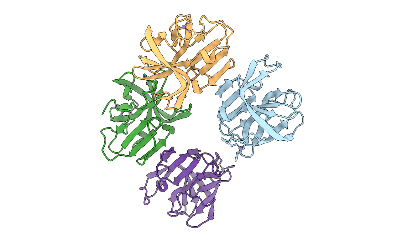
5X45 image
Entry Detail
PDB ID:
5X45
Keywords:
Title:
Crystal structure of 2A protease from Human rhinovirus C15
Biological Source:
Source Organism:
Host Organism:
PDB Version:
Deposition Date:
2017-02-10
Release Date:
2018-02-21
Method Details:
Experimental Method:
Resolution:
2.60 Å
R-Value Free:
0.21
R-Value Work:
0.16
R-Value Observed:
0.16
Space Group:
C 2 2 21
Macromolecular Entities
Polymer Type:polypeptide(L)
Description:protease 2A
Chain IDs:A, B, C, D
Chain Length:139
Number of Molecules:4
Biological Source:Rhinovirus C
Ligand Molecules
Primary Citation
Structural view of the 2A protease from human rhinovirus C15.
Acta Crystallogr.,Sect.F 74 255 261 (2018)
PMID: 29633974 DOI: 10.1107/S2053230X18003382

Abstact

The majority of outbreaks of the common cold are caused by rhinoviruses. The 2A protease (2Apro) of human rhinoviruses (HRVs) is known to play important roles in the propagation of the virus and the modulation of host signal pathways to facilitate viral replication. The 2Apro from human rhinovirus C15 (HRV-C15) has been expressed in Escherichia coli and purified by affinity chromatography, ion-exchange chromatography and gel-filtration chromatography. The crystals diffracted to 2.6 Å resolution. The structure was solved by molecular replacement using the structure of 2Apro from coxsackievirus A16 (CVA16) as the search model. The structure contains a conserved His-Asp-Cys catalytic triad and a Zn2+-binding site. Comparison with other 2Apro structures from enteroviruses reveals that the substrate-binding cleft of 2Apro from HRV-C15 exhibits a more open conformation, which presumably favours substrate binding.

Legend

Protein

Chemical

Disease

Primary Citation of related structures