5X3Z image
Deposition Date 2017-02-09
Release Date 2017-12-13
Last Version Date 2024-05-01
Entry Detail
PDB ID:
5X3Z
Title:
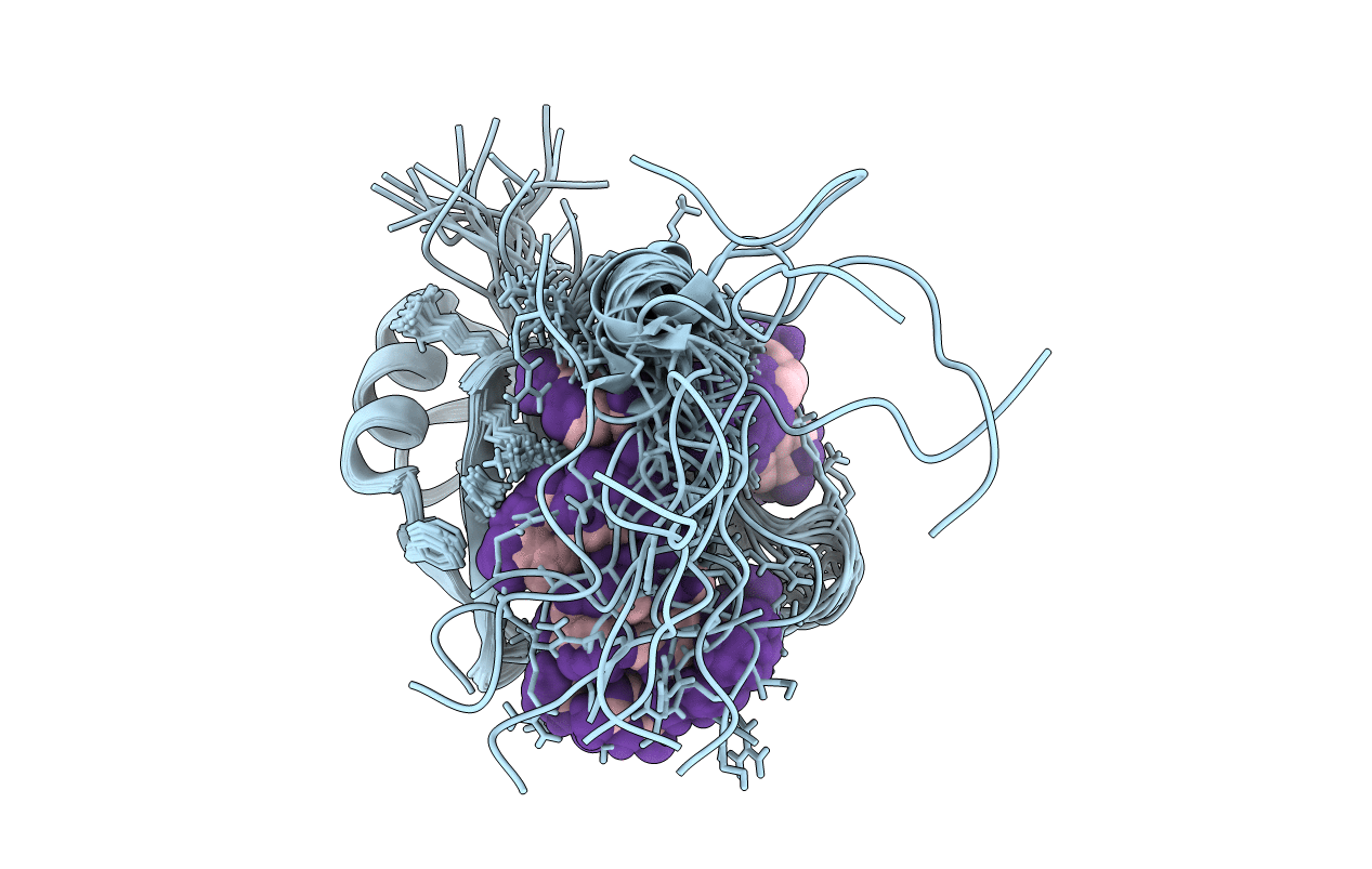
Solution structure of musashi1 RBD2 in complex with RNA
Biological Source:
Source Organism:
Mus musculus (Taxon ID: 10090)
Host Organism:
Method Details:
Experimental Method:
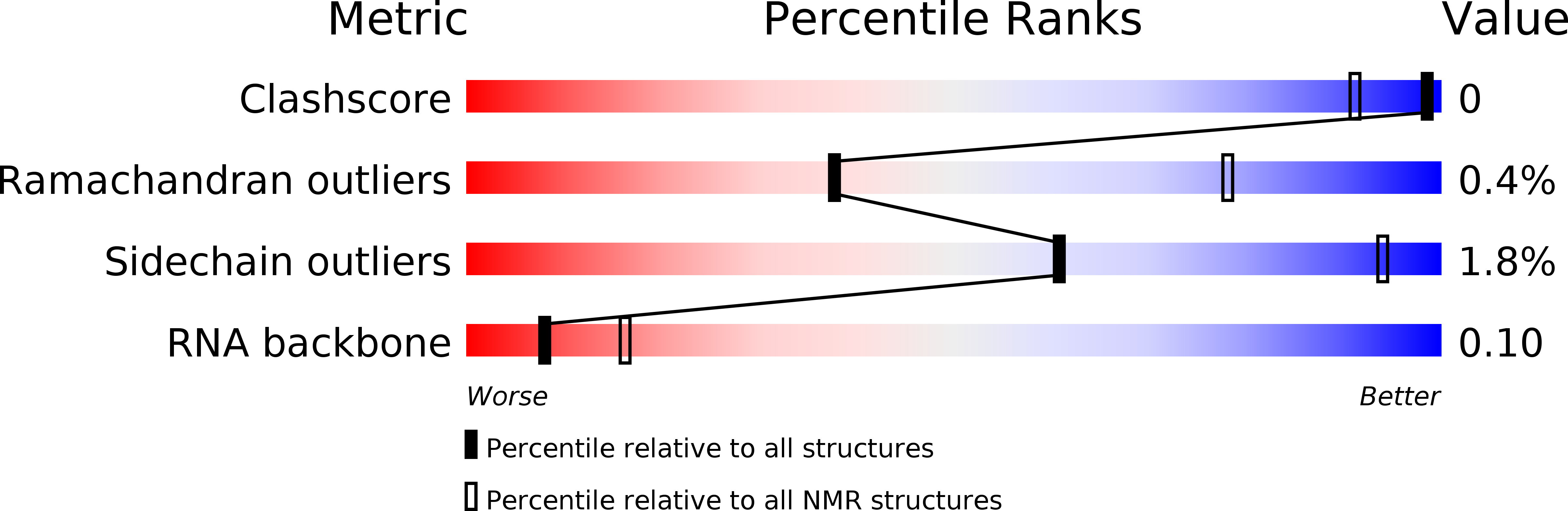
Conformers Calculated:
200
Conformers Submitted:
20
Selection Criteria:
structures with the least restraint violations
Macromolecular Entities
Polymer Type:polypeptide(L)
Molecule:RNA-binding protein Musashi homolog 1
Gene (Uniprot):Msi1
Chain IDs:A
Chain Length:96
Number of Molecules:1
Biological Source:Mus musculus
Polymer Type:polyribonucleotide
Molecule:RNA (5'-R(*GP*UP*AP*GP*U)-3')
Chain IDs:B
Chain Length:5
Number of Molecules:1
Biological Source:Mus musculus
Ligand Molecules
Primary Citation
Structural Insight into the Recognition of r(UAG) by Musashi-1 RBD2, and Construction of a Model of Musashi-1 RBD1-2 Bound to the Minimum Target RNA
Molecules 22 ? ? (2017)
PMID: 28753936 DOI: 10.3390/molecules22071207

Abstact

Musashi-1 (Msi1) controls the maintenance of stem cells and tumorigenesis through binding to its target mRNAs and subsequent translational regulation. Msi1 has two RNA-binding domains (RBDs), RBD1 and RBD2, which recognize r(GUAG) and r(UAG), respectively. These minimal recognition sequences are connected by variable linkers in the Msi1 target mRNAs, however, the molecular mechanism by which Msi1 recognizes its targets is not yet understood. We previously determined the solution structure of the Msi1 RBD1:r(GUAGU) complex. Here, we determined the first structure of the RBD2:r(GUAGU) complex. The structure revealed that the central trinucleotide, r(UAG), is specifically recognized by the intermolecular hydrogen-bonding and aromatic stacking interactions. Importantly, the C-terminal region, which is disordered in the free form, took a certain conformation, resembling a helix. The observation of chemical shift perturbation and intermolecular NOEs, together with increases in the heteronuclear steady-state {¹H}-15N NOE values on complex formation, indicated the involvement of the C-terminal region in RNA binding. On the basis of the two complex structures, we built a structural model of consecutive RBDs with r(UAGGUAG) containing both minimal recognition sequences, which resulted in no steric hindrance. The model suggests recognition of variable lengths (n) of the linker up to n = 50 may be possible.

Legend

Protein

Chemical

Disease

Primary Citation of related structures