5X2J image
Deposition Date 2017-01-31
Release Date 2018-01-31
Last Version Date 2023-11-22
Entry Detail
PDB ID:
5X2J
Keywords:
Title:
Crystal structure of a recombinant hybrid manganese superoxide dismutase from Staphylococcus equorum and Staphylococcus saprophyticus
Biological Source:
Source Organism:
Method Details:
Experimental Method:
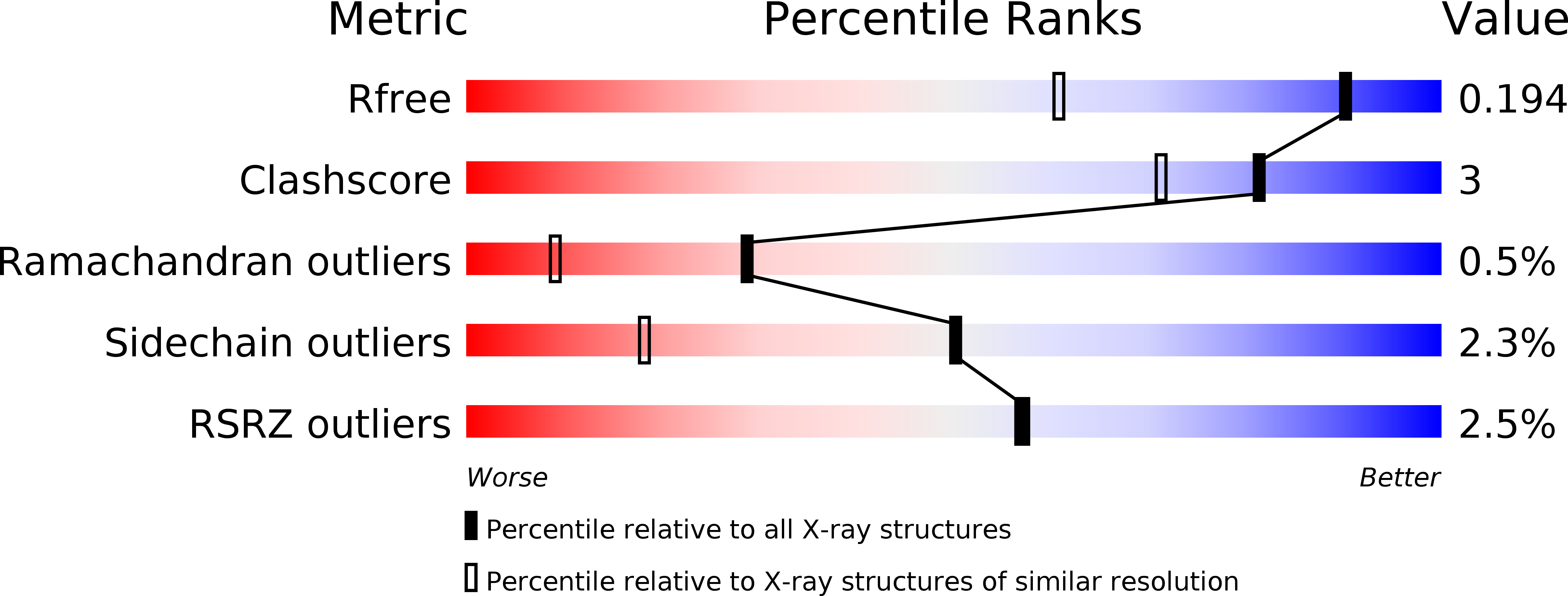
Resolution:
1.40 Å
R-Value Free:
0.18
R-Value Work:
0.14
R-Value Observed:
0.14
Space Group:
P 32 2 1
Macromolecular Entities
Polymer Type:polypeptide(L)
Molecule:manganese superoxide dismutase
Gene (Uniprot):M4L21_03170
Mutations:D13R
Chain IDs:A
Chain Length:199
Number of Molecules:1
Biological Source:Staphylococcus equorum
Ligand Molecules
Primary Citation
The first crystal structure of manganese superoxide dismutase from the genus Staphylococcus
Acta Crystallogr F Struct Biol Commun 74 135 142 (2018)
PMID: 29497016 DOI: 10.1107/S2053230X18001036

Abstact

A recombinant Staphylococcus equorum manganese superoxide dismutase (MnSOD) with an Asp13Arg substitution displays activity over a wide range of pH, at high temperature and in the presence of chaotropic agents, and retains 50% of its activity after irradiation with UVC for up to 45 min. Interestingly, Bacillus subtilis MnSOD does not have the same stability, despite having a closely similar primary structure and thus presumably also tertiary structure. Here, the crystal structure of S. equorum MnSOD at 1.4 Å resolution is reported that may explain these differences. The crystal belonged to space group P3221, with unit-cell parameters a = 57.36, b = 57.36, c = 105.76 Å, and contained one molecule in the asymmetric unit. The symmetry operation indicates that the enzyme has a dimeric structure, as found in nature and in B. subtilis MnSOD. As expected, their overall structures are nearly identical. However, the loop connecting the helical and α/β domains of S. equorum MnSOD is shorter than that in B. subtilis MnSOD, and adopts a conformation that allows more direct water-mediated hydrogen-bond interactions between the amino-acid side chains of the first and last α-helices in the latter domain. Furthermore, S. equorum MnSOD has a slightly larger buried area compared with the dimer surface area than that in B. subtilis MnSOD, while the residues that form the interaction in the dimer-interface region are highly conserved. Thus, the stability of S. equorum MnSOD may not originate from the dimeric form alone. Furthermore, an additional water molecule was found in the active site. This allows an alternative geometry for the coordination of the Mn atom in the active site of the apo form. This is the first structure of MnSOD from the genus Staphylococcus and may provide a template for the structural study of other MnSODs from this genus.

Legend

Protein

Chemical

Disease

Primary Citation of related structures