
Deposition Date
2017-01-26
Release Date
2017-05-17
Last Version Date
2023-11-22
Entry Detail
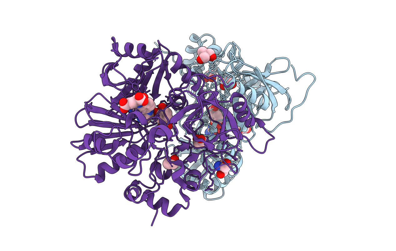
PDB ID:
5X1N
Keywords:
Title:
Vanillate/3-O-methylgallate O-demethylase, LigM, protocatechuate-tetrahydrofolate complex form
Biological Source:
Source Organism(s):
Sphingobium sp. SYK-6 (Taxon ID: 627192)
Expression System(s):
Method Details:
Experimental Method:
Resolution:
2.00 Å
R-Value Free:
0.23
R-Value Work:
0.21
R-Value Observed:
0.21
Space Group:
P 31 2 1
