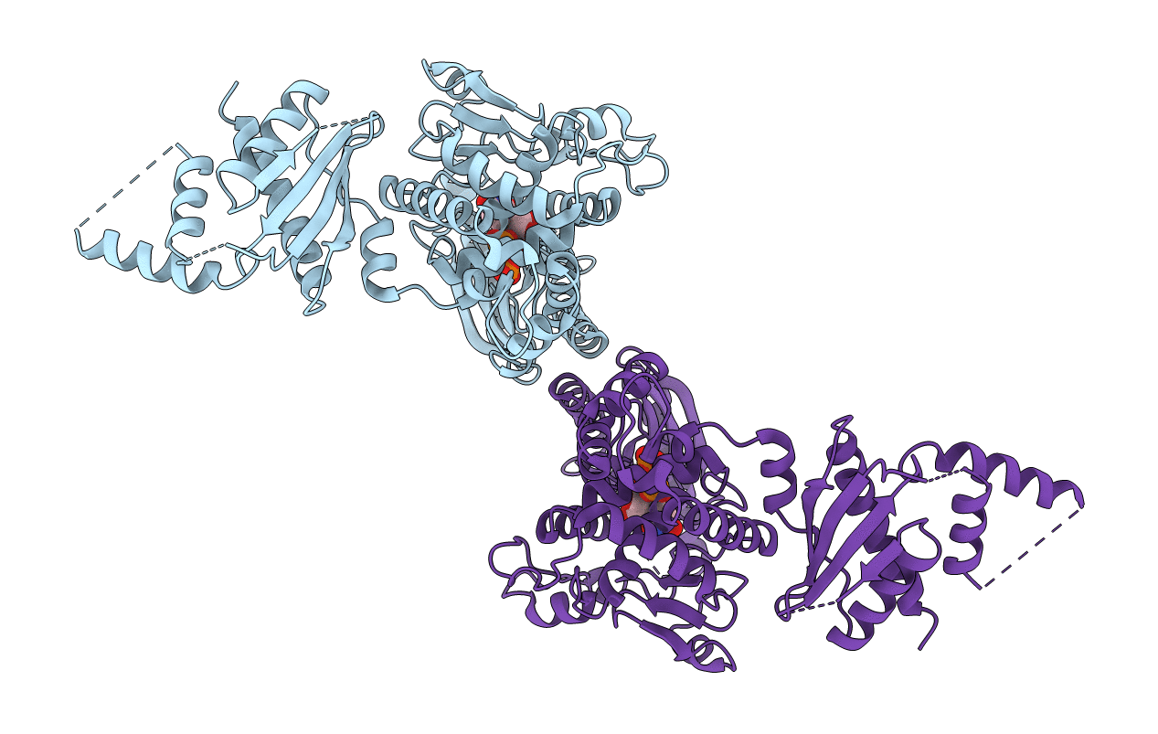
5WU2 image
Entry Detail
PDB ID:
5WU2
Keywords:
Title:
Crystal structure of human Tut1 bound with BaUTP, form I
Biological Source:
Source Organism:
Host Organism:
PDB Version:
Deposition Date:
2016-12-16
Release Date:
2017-05-31
Method Details:
Experimental Method:
Resolution:
2.95 Å
R-Value Free:
0.29
R-Value Work:
0.24
R-Value Observed:
0.24
Space Group:
P 21 21 21
Macromolecular Entities
Polymer Type:polypeptide(L)
Description:Speckle targeted PIP5K1A-regulated poly(A) polymerase
Mutations:C372A, C415A, C501A, C504S
Chain IDs:A, B
Chain Length:573
Number of Molecules:2
Biological Source:Homo sapiens
Primary Citation
Crystal structures of U6 snRNA-specific terminal uridylyltransferase
Nat Commun 8 15788 15788 (2017)
PMID: 28589955 DOI: 10.1038/ncomms15788

Abstact

The terminal uridylyltransferase, TUT1, builds or repairs the 3'-oligo-uridylylated tail of U6 snRNA. The 3'-oligo-uridylylated tail is the Lsm-binding site for U4/U6 di-snRNP formation and U6 snRNA recycling for pre-mRNA splicing. Here, we report crystallographic and biochemical analyses of human TUT1, which revealed the mechanisms for the specific uridylylation of the 3'-end of U6 snRNA by TUT1. The O2 and O4 atoms of the UTP base form hydrogen bonds with the conserved His and Asn in the catalytic pocket, respectively, and TUT1 preferentially incorporates UMP onto the 3'-end of RNAs. TUT1 recognizes the entire U6 snRNA molecule by its catalytic domains, N-terminal RNA-recognition motifs and a previously unidentified C-terminal RNA-binding domain. Each domain recognizes specific regions within U6 snRNA, and the recognition is coupled with the domain movements and U6 snRNA structural changes. Hence, TUT1 functions as the U6 snRNA-specific terminal uridylyltransferase required for pre-mRNA splicing.

Legend

Protein

Chemical

Disease

Primary Citation of related structures