5WTN image
Deposition Date 2016-12-13
Release Date 2017-08-23
Last Version Date 2024-03-20
Entry Detail
PDB ID:
5WTN
Keywords:
Title:
Crystal Structure Analysis of primosome protein DnaB (resiues 1-300) from Geobacillus stearothermophilus
Biological Source:
Source Organism:
Host Organism:
Method Details:
Experimental Method:
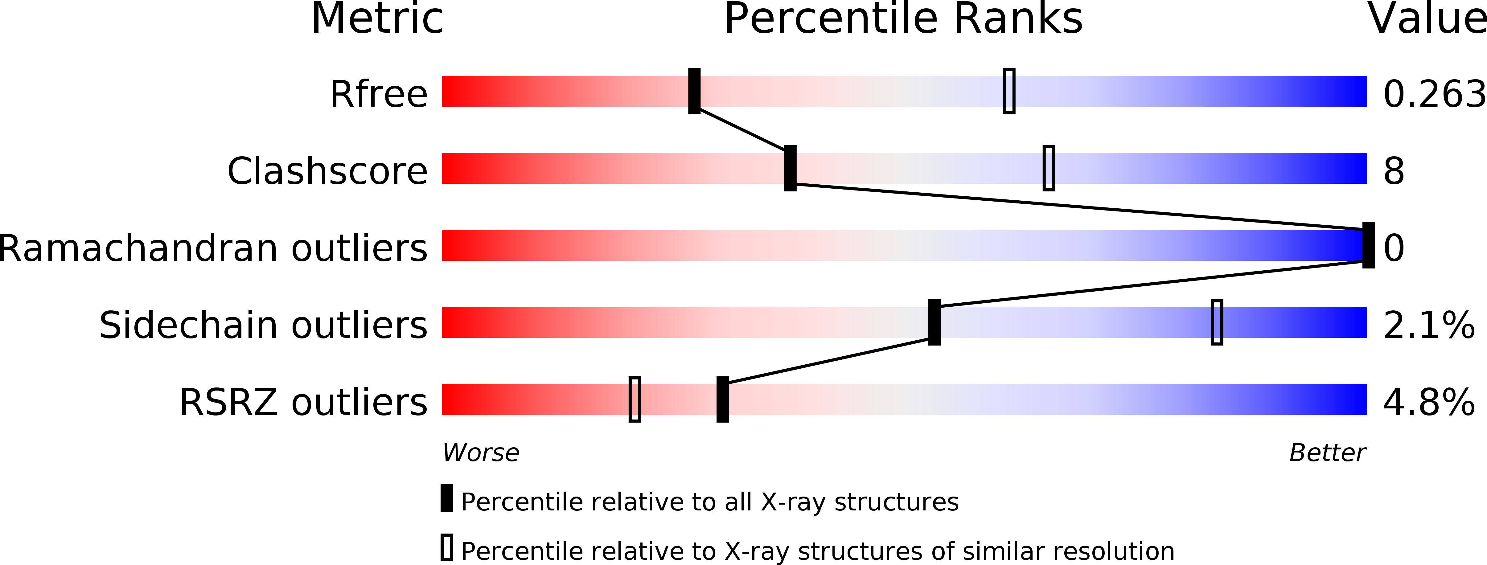
Resolution:
2.80 Å
R-Value Free:
0.26
R-Value Work:
0.22
R-Value Observed:
0.22
Space Group:
P 21 21 21
Macromolecular Entities
Polymer Type:polypeptide(L)
Molecule:Replication initiation and membrane attachment protein
Chain IDs:A, B, C, D
Chain Length:300
Number of Molecules:4
Biological Source:Geobacillus stearothermophilus 10
Primary Citation
Structural analyses of the bacterial primosomal protein DnaB reveal that it is a tetramer and forms a complex with a primosomal re-initiation protein
J. Biol. Chem. 292 15744 15757 (2017)
PMID: 28808061 DOI: 10.1074/jbc.M117.792002

Abstact

The DnaB primosomal protein from Gram-positive bacteria plays a key role in DNA replication and restart as a loader protein for the recruitment of replisome cascade proteins. Previous investigations have established that DnaB is composed of an N-terminal domain, a middle domain, and a C-terminal domain. However, structural evidence for how DnaB functions at the atomic level is lacking. Here, we report the crystal structure of DnaB, encompassing the N-terminal and middle domains (residues 1-300), from Geobacillus stearothermophilus (GstDnaB1-300) at 2.8 Å resolution. Our structure revealed that GstDnaB1-300 forms a tetramer with two basket-like architectures, a finding consistent with those from solution studies using analytical ultracentrifugation. Furthermore, our results from both GST pulldown assays and analytical ultracentrifugation show that GstDnaB1-300 is sufficient to form a complex with PriA, the primosomal reinitiation protein. Moreover, with the aid of small angle X-ray scattering experiments, we also determined the structural envelope of full-length DnaB (GstDnaBFL) in solution. These small angle X-ray scattering studies indicated that GstDnaBFL has an elongated conformation and that the protruding density envelopes originating from GstDnaB1-300 could completely accommodate the GstDnaB C-terminal domain (residues 301-461). Taken together with biochemical assays, our results suggest that GstDnaB uses different domains to distinguish the PriA interaction and single-stranded DNA binding. These findings can further extend our understanding of primosomal assembly in replication restart.

Legend

Protein

Chemical

Disease

Primary Citation of related structures