5WS6 image
Entry Detail
PDB ID:
5WS6
Keywords:
Title:
Native XFEL structure of Photosystem II (preflash two-flash dataset
Biological Source:
PDB Version:
Deposition Date:
2016-12-05
Release Date:
2017-03-15
Method Details:
Experimental Method:
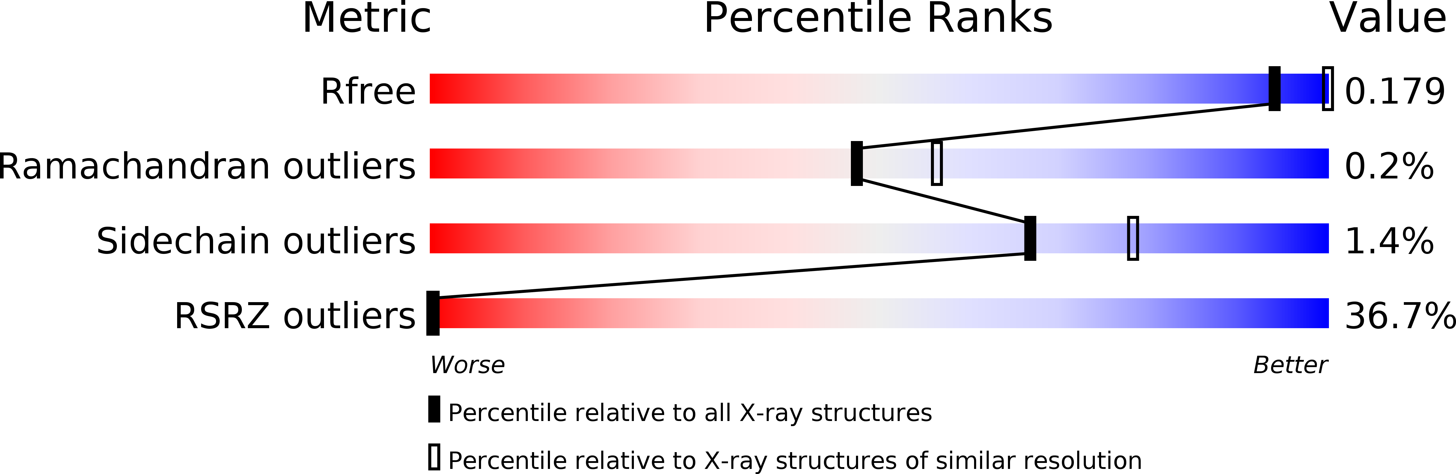
Resolution:
2.35 Å
R-Value Free:
0.17
R-Value Work:
0.12
R-Value Observed:
0.13
Space Group:
P 21 21 21
Macromolecular Entities
Polymer Type:polypeptide(L)
Description:Photosystem II D1 protein
Chain IDs:A, U (auth: a)
Chain Length:344
Number of Molecules:2
Biological Source:Thermosynechococcus vulcanus
Polymer Type:polypeptide(L)
Description:Photosystem II CP47 reaction center protein
Chain IDs:B, V (auth: b)
Chain Length:505
Number of Molecules:2
Biological Source:Thermosynechococcus vulcanus
Polymer Type:polypeptide(L)
Description:Photosystem II CP43 chlorophyll protein
Chain IDs:C, W (auth: c)
Chain Length:455
Number of Molecules:2
Biological Source:Thermosynechococcus vulcanus
Polymer Type:polypeptide(L)
Description:Photosystem II D2 protein
Chain IDs:D, X (auth: d)
Chain Length:342
Number of Molecules:2
Biological Source:Thermosynechococcus vulcanus
Polymer Type:polypeptide(L)
Description:Cytochrome b559 subunit alpha
Chain IDs:E, Y (auth: e)
Chain Length:84
Number of Molecules:2
Biological Source:Thermosynechococcus vulcanus
Polymer Type:polypeptide(L)
Description:Cytochrome b559 subunit beta
Chain IDs:F, Z (auth: f)
Chain Length:44
Number of Molecules:2
Biological Source:Thermosynechococcus vulcanus
Polymer Type:polypeptide(L)
Description:Photosystem II reaction center protein H
Chain IDs:G (auth: H), AA (auth: h)
Chain Length:65
Number of Molecules:2
Biological Source:Thermosynechococcus vulcanus
Polymer Type:polypeptide(L)
Description:Photosystem II reaction center protein I
Chain IDs:H (auth: I), BA (auth: i)
Chain Length:38
Number of Molecules:2
Biological Source:Thermosynechococcus vulcanus
Polymer Type:polypeptide(L)
Description:Photosystem II reaction center protein J
Chain IDs:I (auth: J), CA (auth: j)
Chain Length:39
Number of Molecules:2
Biological Source:Thermosynechococcus vulcanus
Polymer Type:polypeptide(L)
Description:Photosystem II reaction center protein K
Chain IDs:J (auth: K), DA (auth: k)
Chain Length:37
Number of Molecules:2
Biological Source:Thermosynechococcus vulcanus
Polymer Type:polypeptide(L)
Description:Photosystem II reaction center protein L
Chain IDs:K (auth: L), EA (auth: l)
Chain Length:37
Number of Molecules:2
Biological Source:Thermosynechococcus vulcanus
Polymer Type:polypeptide(L)
Description:Photosystem II reaction center protein M
Chain IDs:L (auth: M), FA (auth: m)
Chain Length:36
Number of Molecules:2
Biological Source:Thermosynechococcus vulcanus
Polymer Type:polypeptide(L)
Description:Photosystem II manganese-stabilizing polypeptide
Chain IDs:M (auth: O), GA (auth: o)
Chain Length:244
Number of Molecules:2
Biological Source:Thermosynechococcus vulcanus
Polymer Type:polypeptide(L)
Description:Photosystem II protein Y
Chain IDs:T (auth: R)
Chain Length:34
Number of Molecules:1
Biological Source:Thermosynechococcus vulcanus
Polymer Type:polypeptide(L)
Description:Photosystem II reaction center protein T
Chain IDs:N (auth: T), HA (auth: t)
Chain Length:32
Number of Molecules:2
Biological Source:Thermosynechococcus vulcanus
Polymer Type:polypeptide(L)
Description:Photosystem II 12 kDa extrinsic protein
Chain IDs:O (auth: U), IA (auth: u)
Chain Length:104
Number of Molecules:2
Biological Source:Thermosynechococcus vulcanus
Polymer Type:polypeptide(L)
Description:Cytochrome c-550
Chain IDs:P (auth: V), JA (auth: v)
Chain Length:137
Number of Molecules:2
Biological Source:Thermosynechococcus vulcanus
Polymer Type:polypeptide(L)
Description:Photosystem II reaction center protein X
Chain IDs:Q (auth: X), KA (auth: x)
Chain Length:40
Number of Molecules:2
Biological Source:Thermosynechococcus vulcanus
Polymer Type:polypeptide(L)
Description:Photosystem II reaction center protein Ycf12
Chain IDs:R (auth: Y), LA (auth: y)
Chain Length:30
Number of Molecules:2
Biological Source:Thermosynechococcus vulcanus
Polymer Type:polypeptide(L)
Description:Photosystem II reaction center protein Z
Chain IDs:S (auth: Z), MA (auth: z)
Chain Length:62
Number of Molecules:2
Biological Source:Thermosynechococcus vulcanus
Modified Residue
Compound ID Chain ID Parent Comp ID Details 2D Image
FME H MET modified residue
Primary Citation

Abstact

Photosystem II (PSII) is a huge membrane-protein complex consisting of 20 different subunits with a total molecular mass of 350 kDa for a monomer. It catalyses light-driven water oxidation at its catalytic centre, the oxygen-evolving complex (OEC). The structure of PSII has been analysed at 1.9 Å resolution by synchrotron radiation X-rays, which revealed that the OEC is a Mn4CaO5 cluster organized in an asymmetric, 'distorted-chair' form. This structure was further analysed with femtosecond X-ray free electron lasers (XFEL), providing the 'radiation damage-free' structure. The mechanism of O=O bond formation, however, remains obscure owing to the lack of intermediate-state structures. Here we describe the structural changes in PSII induced by two-flash illumination at room temperature at a resolution of 2.35 Å using time-resolved serial femtosecond crystallography with an XFEL provided by the SPring-8 ångström compact free-electron laser. An isomorphous difference Fourier map between the two-flash and dark-adapted states revealed two areas of apparent changes: around the QB/non-haem iron and the Mn4CaO5 cluster. The changes around the QB/non-haem iron region reflected the electron and proton transfers induced by the two-flash illumination. In the region around the OEC, a water molecule located 3.5 Å from the Mn4CaO5 cluster disappeared from the map upon two-flash illumination. This reduced the distance between another water molecule and the oxygen atom O4, suggesting that proton transfer also occurred. Importantly, the two-flash-minus-dark isomorphous difference Fourier map showed an apparent positive peak around O5, a unique μ4-oxo-bridge located in the quasi-centre of Mn1 and Mn4 (refs 4,5). This suggests the insertion of a new oxygen atom (O6) close to O5, providing an O=O distance of 1.5 Å between these two oxygen atoms. This provides a mechanism for the O=O bond formation consistent with that proposed previously.

Legend

Protein

Chemical

Disease

Primary Citation of related structures