Deposition Date
2016-11-29
Release Date
2016-12-07
Last Version Date
2024-03-20
Entry Detail
PDB ID:
5WQW
Keywords:
Title:
X-ray structure of catalytic domain of autolysin from Clostridium perfringens
Biological Source:
Source Organism(s):
Clostridium perfringens (strain 13 / Type A) (Taxon ID: 195102)
Expression System(s):
Method Details:
Experimental Method:
Resolution:
1.76 Å
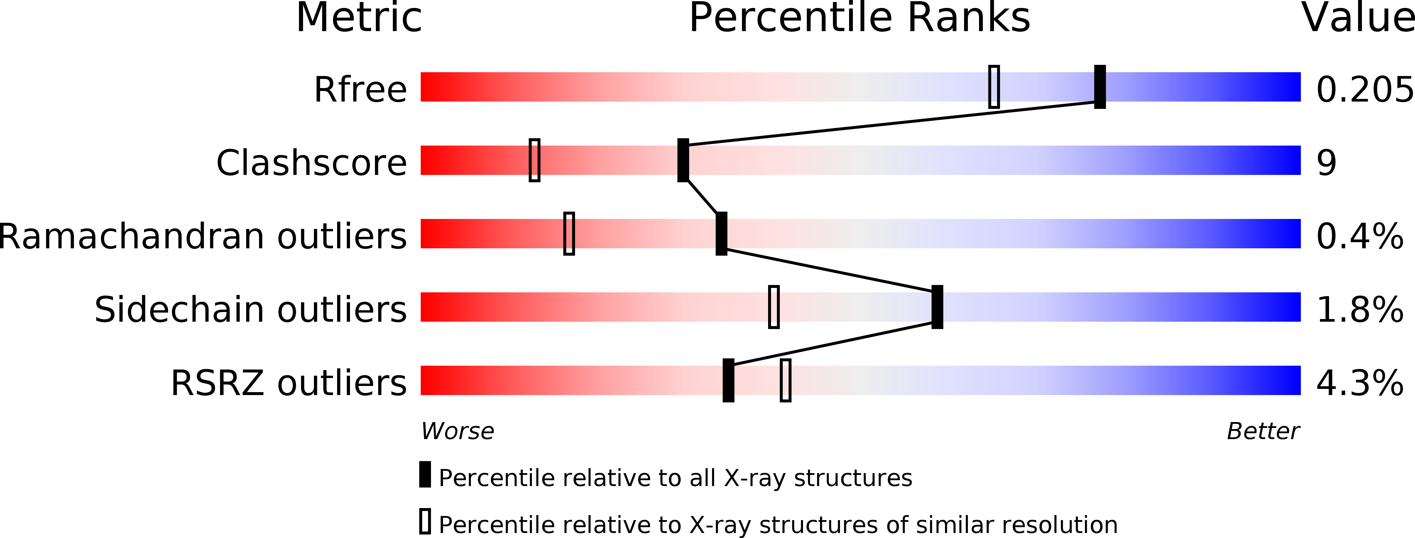
R-Value Free:
0.19
R-Value Work:
0.16
R-Value Observed:
0.16
Space Group:
P 1 21 1