5WQ0 image
Deposition Date 2016-11-22
Release Date 2017-03-22
Last Version Date 2024-03-20
Entry Detail
PDB ID:
5WQ0
Title:
Receiver domain of Spo0A from Paenisporosarcina sp. TG-14
Biological Source:
Source Organism:
Host Organism:
Method Details:
Experimental Method:
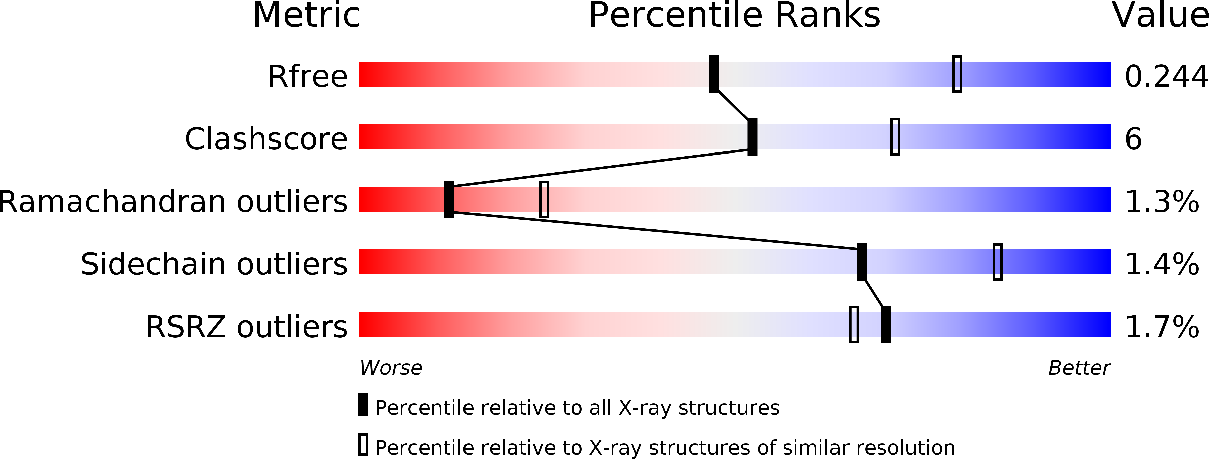
Resolution:
2.60 Å
R-Value Free:
0.24
R-Value Work:
0.18
R-Value Observed:
0.18
Space Group:
P 21 21 21
Macromolecular Entities
Structures with similar UniProt ID
Protein Blast
Polymer Type:polypeptide(L)
Molecule:Stage 0 sporulation protein
Chain IDs:A (auth: B), B (auth: A), C, E, F, G
Chain Length:144
Number of Molecules:6
Biological Source:Paenisporosarcina sp. TG-14
Structures with similar UniProt ID
Protein Blast
Polymer Type:polypeptide(L)
Molecule:Stage 0 sporulation protein
Chain IDs:D
Chain Length:148
Number of Molecules:1
Biological Source:Paenisporosarcina sp. TG-14
Ligand Molecules
Primary Citation
Crystal structure of the inactive state of the receiver domain of Spo0A from Paenisporosarcina sp. TG-14, a psychrophilic bacterium isolated from an Antarctic glacier
J. Microbiol. 55 464 474 (2017)
PMID: 28281198 DOI: 10.1007/s12275-017-6599-9

Abstact

The two-component phosphorelay system is the most prevalent mechanism for sensing and transducing environmental signals in bacteria. Spore formation, which relies on the two-component phosphorelay system, enables the long-term survival of the glacial bacterium Paenisporosarcina sp. TG-14 in the extreme cold environment. Spo0A is a key response regulator of the phosphorelay system in the early stage of spore formation. The protein is composed of a regulatory N-terminal phospho-receiver domain and a DNA-binding C-terminal activator domain. We solved the three-dimensional structure of the unphosphorylated (inactive) form of the receiver domain of Spo0A (PaSpo0A-R) from Paenisporosarcina sp. TG-14. A structural comparison with phosphorylated (active form) Spo0A from Bacillus stearothermophilus (BsSpo0A) showed minor notable differences. A molecular dynamics study of a model of the active form and the crystal structures revealed significant differences in the α4 helix and the preceding loop region where phosphorylation occurs. Although an oligomerization study of PaSpo0A-R by analytical ultracentrifugation (AUC) has shown that the protein is in a monomeric state in solution, both crosslinking and crystal-packing analyses indicate the possibility of weak dimer formation by a previously undocumented mechanism. Collectively, these observations provide insight into the mechanism of phosphorylation-dependent activation unique to Spo0A.

Legend

Protein

Chemical

Disease

Primary Citation of related structures
Feedback Form
Name
Email
Institute
Feedback