5WOU image
Deposition Date 2017-08-03
Release Date 2018-02-28
Last Version Date 2024-11-13
Entry Detail
PDB ID:
5WOU
Keywords:
Title:
Crystal Structure of drosophila melanogaster Scribble PDZ1 domain in complex with Guk-Holder
Biological Source:
Source Organism:
Host Organism:
Method Details:
Experimental Method:
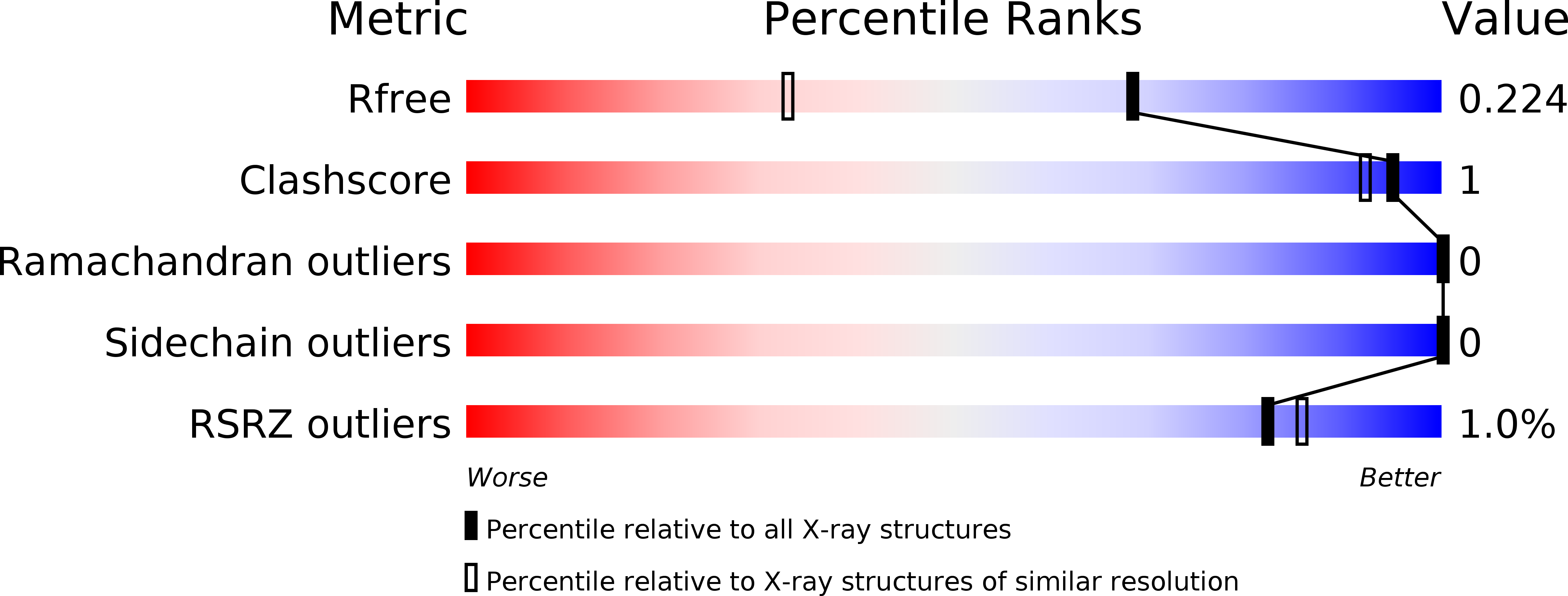
Resolution:
1.55 Å
R-Value Free:
0.22
R-Value Work:
0.19
R-Value Observed:
0.19
Space Group:
C 2 2 21
Macromolecular Entities
Structures with similar UniProt ID
Protein Blast
Polymer Type:polypeptide(L)
Molecule:Protein lap4
Gene (Uniprot):scrib
Chain IDs:A
Chain Length:100
Number of Molecules:1
Biological Source:Drosophila melanogaster
Structures with similar UniProt ID
Protein Blast
Polymer Type:polypeptide(L)
Molecule:GUK-holder, isoform A
Gene (Uniprot):gukh
Chain IDs:B (auth: V)
Chain Length:8
Number of Molecules:1
Biological Source:Drosophila melanogaster
Primary Citation
Drosophila melanogasterGuk-holder interacts with the Scribbled PDZ1 domain and regulates epithelial development with Scribbled and Discs Large.
J. Biol. Chem. 293 4519 4531 (2018)
PMID: 29378849 DOI: 10.1074/jbc.M117.817528

Abstact

Epithelial cell polarity is controlled by components of the Scribble polarity module, and its regulation is critical for tissue architecture and cell proliferation and migration. In Drosophila melanogaster, the adaptor protein Guk-holder (Gukh) binds to the Scribbled (Scrib) and Discs Large (Dlg) components of the Scribble polarity module and plays an important role in the formation of neuromuscular junctions. However, Gukh's role in epithelial tissue formation and the molecular basis for the Scrib-Gukh interaction remain to be defined. We now show using isothermal titration calorimetry that the Scrib PDZ1 domain is the major site for an interaction with Gukh. Furthermore, we defined the structural basis of this interaction by determining the crystal structure of the Scrib PDZ1-Gukh complex. The C-terminal PDZ-binding motif of Gukh is located in the canonical ligand-binding groove of Scrib PDZ1 and utilizes an unusually extensive network of hydrogen bonds and ionic interactions to enable binding to PDZ1 with high affinity. We next examined the role of Gukh along with those of Scrib and Dlg in Drosophila epithelial tissues and found that Gukh is expressed in larval-wing and eye-epithelial tissues and co-localizes with Scrib and Dlg at the apical cell cortex. Importantly, we show that Gukh functions with Scrib and Dlg in the development of Drosophila epithelial tissues, with depletion of Gukh enhancing the eye- and wing-tissue defects caused by Scrib or Dlg depletion. Overall, our findings reveal that Scrib's PDZ1 domain functions in the interaction with Gukh and that the Scrib-Gukh interaction has a key role in epithelial tissue development in Drosophila.

Legend

Protein

Chemical

Disease

Primary Citation of related structures