5WOB image
Deposition Date 2017-08-01
Release Date 2018-04-18
Last Version Date 2024-10-23
Entry Detail
PDB ID:
5WOB
Keywords:
Title:
Crystal Structure Analysis of Fab1-Bound Human Insulin Degrading Enzyme (IDE) in Complex with Insulin
Biological Source:
Source Organism:
Homo sapiens (Taxon ID: 9606)
Mus musculoides (Taxon ID: 60742)
Host Organism:
Method Details:
Experimental Method:
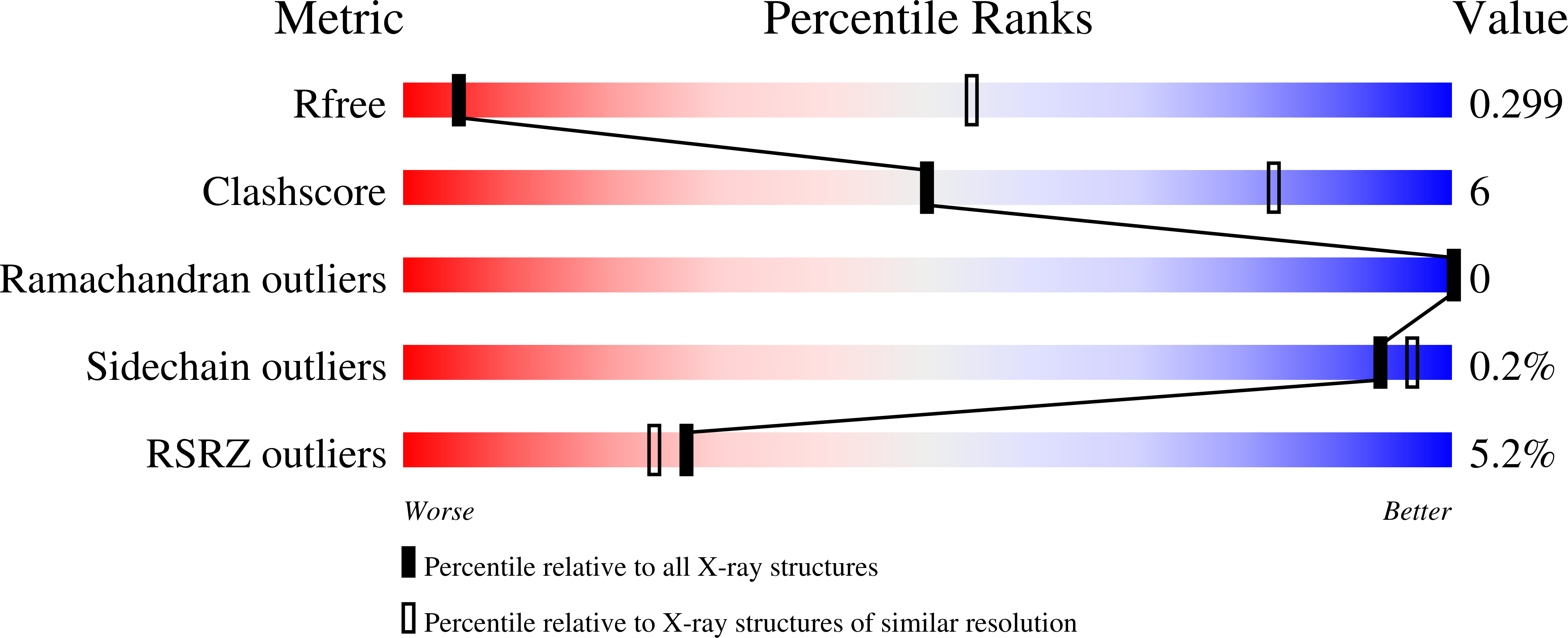
Resolution:
3.95 Å
R-Value Free:
0.29
R-Value Work:
0.24
R-Value Observed:
0.24
Space Group:
P 1 21 1
Macromolecular Entities
Structures with similar UniProt ID
Protein Blast
Polymer Type:polypeptide(L)
Molecule:Insulin-degrading enzyme
Gene (Uniprot):IDE
Chain IDs:A, B, C, D, E, F, G, H
Chain Length:20
Number of Molecules:8
Biological Source:Homo sapiens
Polymer Type:polypeptide(L)
Molecule:IDE-bound Fab heavy chain
Chain IDs:Q (auth: I), R (auth: K), S (auth: M), T (auth: O), U (auth: Q), V (auth: S), W (auth: U), X (auth: W)
Chain Length:263
Number of Molecules:8
Biological Source:Mus musculoides
Polymer Type:polypeptide(L)
Molecule:IDE-bound Fab light chain
Chain IDs:Y (auth: J), Z (auth: L), AA (auth: N), BA (auth: P), CA (auth: R), DA (auth: T), EA (auth: V), FA (auth: X)
Chain Length:239
Number of Molecules:8
Biological Source:Mus musculoides
Structures with similar UniProt ID
Protein Blast
Polymer Type:polypeptide(L)
Molecule:Insulin
Gene (Uniprot):INS
Chain IDs:I (auth: a), J (auth: b), K (auth: c), L (auth: d), M (auth: e), N (auth: f), O (auth: g), P (auth: h)
Chain Length:20
Number of Molecules:8
Biological Source:Homo sapiens
Ligand Molecules
Primary Citation
Ensemble cryoEM elucidates the mechanism of insulin capture and degradation by human insulin degrading enzyme.
Elife 7 ? ? (2018)
PMID: 29596046 DOI: 10.7554/eLife.33572

Abstact

Insulin degrading enzyme (IDE) plays key roles in degrading peptides vital in type two diabetes, Alzheimer's, inflammation, and other human diseases. However, the process through which IDE recognizes peptides that tend to form amyloid fibrils remained unsolved. We used cryoEM to understand both the apo- and insulin-bound dimeric IDE states, revealing that IDE displays a large opening between the homologous ~55 kDa N- and C-terminal halves to allow selective substrate capture based on size and charge complementarity. We also used cryoEM, X-ray crystallography, SAXS, and HDX-MS to elucidate the molecular basis of how amyloidogenic peptides stabilize the disordered IDE catalytic cleft, thereby inducing selective degradation by substrate-assisted catalysis. Furthermore, our insulin-bound IDE structures explain how IDE processively degrades insulin by stochastically cutting either chain without breaking disulfide bonds. Together, our studies provide a mechanism for how IDE selectively degrades amyloidogenic peptides and offers structural insights for developing IDE-based therapies.

Legend

Protein

Chemical

Disease

Primary Citation of related structures
Feedback Form
Name
Email
Institute
Feedback