
Deposition Date
2017-08-01
Release Date
2017-08-16
Last Version Date
2023-10-04
Entry Detail
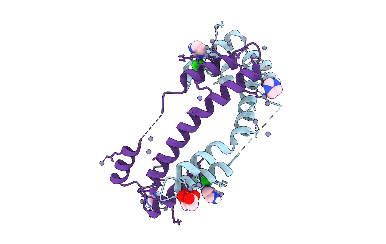
PDB ID:
5WO1
Keywords:
Title:
Chaperone Spy H96L bound to Im7 L18A L19A L37A (Im7 un-modeled)
Biological Source:
Source Organism:
Escherichia coli (Taxon ID: 562)
Host Organism:
Method Details:
Experimental Method:
Resolution:
1.87 Å
R-Value Free:
0.24
R-Value Work:
0.20
R-Value Observed:
0.21
Space Group:
P 41 2 2
