5WHG image
Deposition Date 2017-07-17
Release Date 2017-11-15
Last Version Date 2024-11-20
Entry Detail
PDB ID:
5WHG
Title:
Vms1 mitochondrial localization core
Biological Source:
Method Details:
Experimental Method:
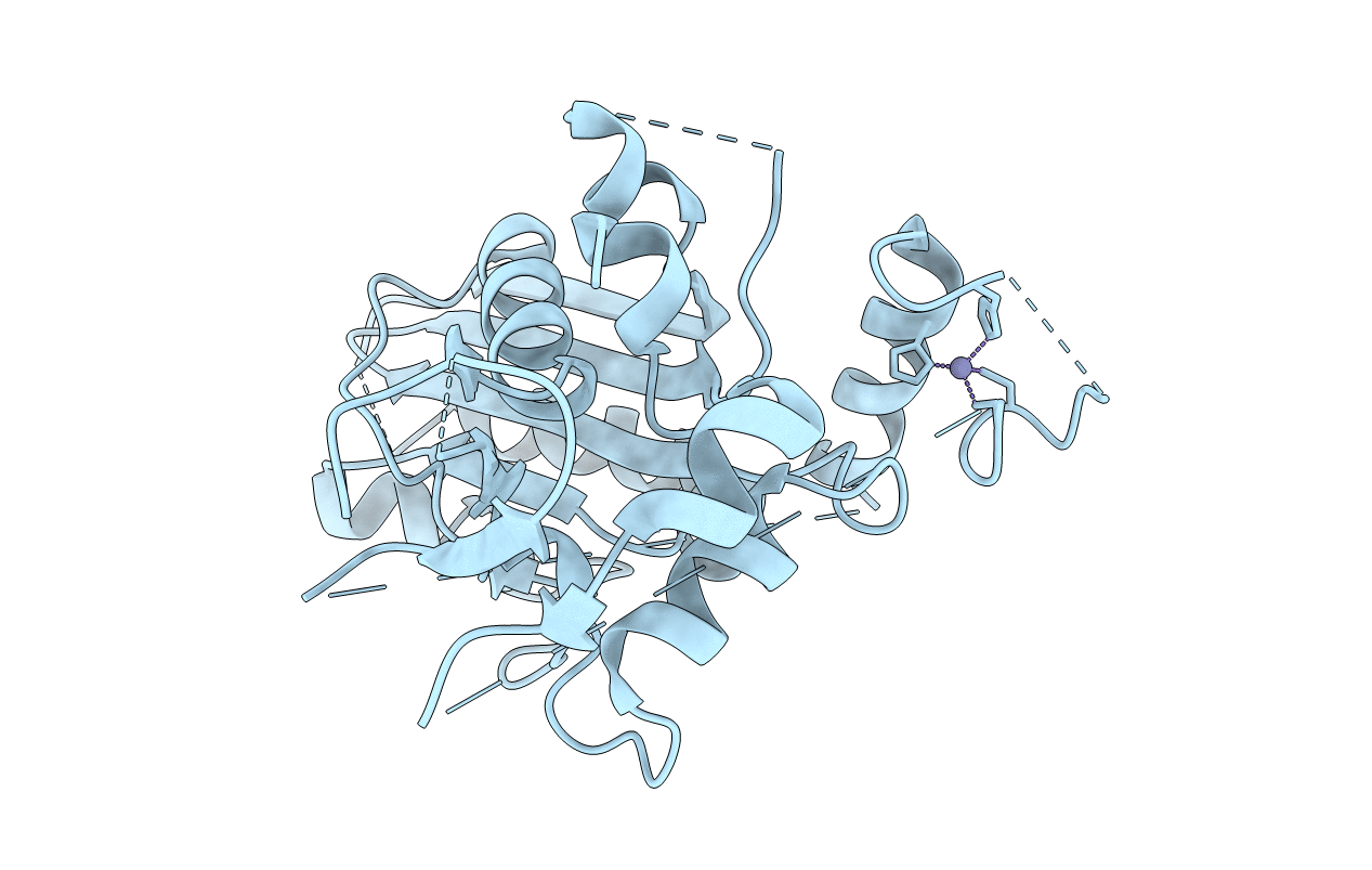
Resolution:
2.70 Å
R-Value Free:
0.24
R-Value Work:
0.18
R-Value Observed:
0.19
Space Group:
P 32 2 1
Macromolecular Entities
Structures with similar UniProt ID
Protein Blast
Polymer Type:polypeptide(L)
Molecule:Protein VMS1
Gene (Uniprot):VMS1
Mutagens:delta 38-69
Chain IDs:A
Chain Length:389
Number of Molecules:1
Biological Source:Saccharomyces cerevisiae (strain ATCC 204508 / S288c)
Modified Residue
Compound ID Chain ID Parent Comp ID Details 2D Image
MSE A MET modified residue
Ligand Molecules
Primary Citation
Sterol Oxidation Mediates Stress-Responsive Vms1 Translocation to Mitochondria.
Mol. Cell 68 673 685.e6 (2017)
PMID: 29149595 DOI: 10.1016/j.molcel.2017.10.022

Abstact

Vms1 translocates to damaged mitochondria in response to stress, whereupon its binding partner, Cdc48, contributes to mitochondrial protein homeostasis. Mitochondrial targeting of Vms1 is mediated by its conserved mitochondrial targeting domain (MTD), which, in unstressed conditions, is inhibited by intramolecular binding to the Vms1 leucine-rich sequence (LRS). Here, we report a 2.7 Å crystal structure of Vms1 that reveals that the LRS lies in a hydrophobic groove in the autoinhibited MTD. We also demonstrate that the oxidized sterol, ergosterol peroxide, is necessary and sufficient for Vms1 localization to mitochondria, through binding the MTD in an interaction that is competitive with binding to the LRS. These data support a model in which stressed mitochondria generate an oxidized sterol receptor that recruits Vms1 to support mitochondrial protein homeostasis.

Legend

Protein

Chemical

Disease

Primary Citation of related structures
Feedback Form
Name
Email
Institute
Feedback