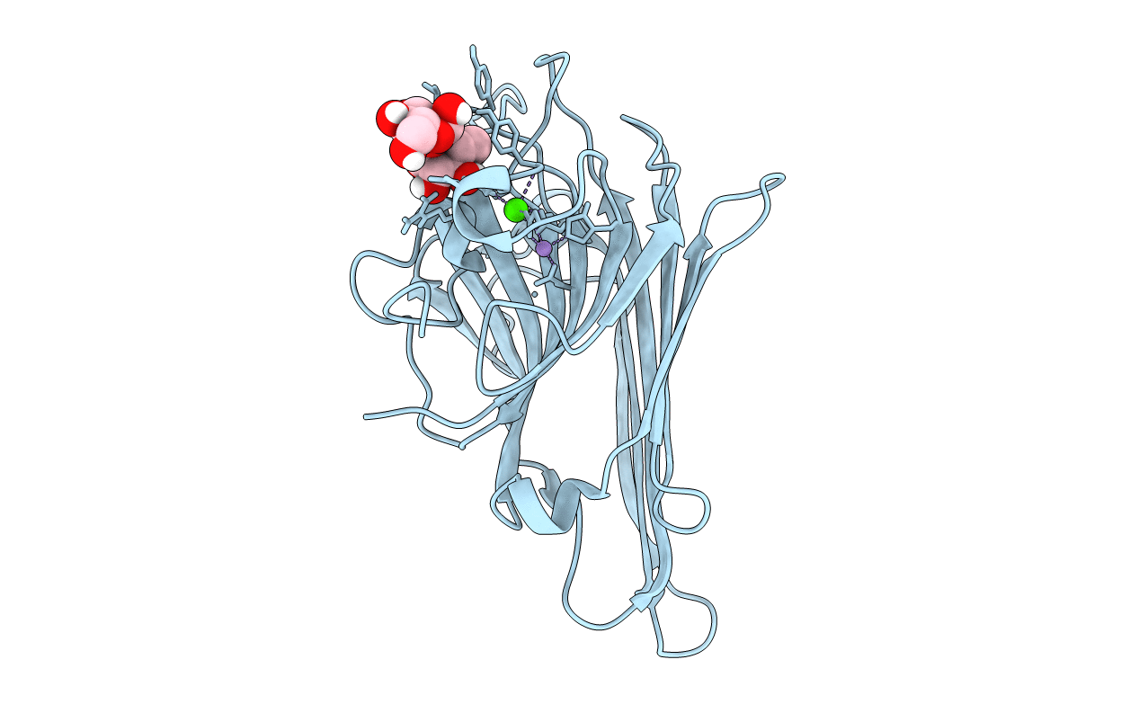
5WEY image
Entry Detail
PDB ID:
5WEY
Title:
Joint X-ray/neutron structure of Concanavalin A with alpha1-2 D-mannobiose
Biological Source:
Source Organism:
PDB Version:
Deposition Date:
2017-07-11
Release Date:
2017-09-13
Method Details:
R-Value Free:
['0.21
R-Value Work:
['0.19
R-Value Observed:
['?', '?'].00
Space Group:
I 2 2 2
Macromolecular Entities
Polymer Type:polypeptide(L)
Description:Concanavalin-A
Chain IDs:A
Chain Length:237
Number of Molecules:1
Biological Source:Canavalia ensiformis
Peptide-like Molecules
PRD_900111
Primary Citation
Mannobiose Binding Induces Changes in Hydrogen Bonding and Protonation States of Acidic Residues in Concanavalin A As Revealed by Neutron Crystallography.
Biochemistry 56 4747 4750 (2017)
PMID: 28846383 DOI: 10.1021/acs.biochem.7b00654

Abstact

Plant lectins are carbohydrate-binding proteins with various biomedical applications. Concanavalin A (Con A) holds promise in treating cancerous tumors. To better understand the Con A carbohydrate binding specificity, we obtained a room-temperature neutron structure of this legume lectin in complex with a disaccharide Manα1-2Man, mannobiose. The neutron structure afforded direct visualization of the hydrogen bonding between the protein and ligand, showing that the ligand is able to alter both protonation states and interactions for residues located close to and distant from the binding site. An unprecedented low-barrier hydrogen bond was observed forming between the carboxylic side chains of Asp28 and Glu8, with the D atom positioned equidistant from the oxygen atoms having an O···D···O angle of 101.5°.

Legend

Protein

Chemical

Disease

Primary Citation of related structures