Deposition Date
2017-07-02
Release Date
2018-10-31
Last Version Date
2023-10-04
Entry Detail
PDB ID:
5WCU
Keywords:
Title:
Crystal structure of 167 bp nucleosome bound to the globular domain of linker histone H5
Biological Source:
Source Organism(s):
Drosophila melanogaster (Taxon ID: 7227)
Gallus gallus (Taxon ID: 9031)
synthetic construct (Taxon ID: 32630)
Gallus gallus (Taxon ID: 9031)
synthetic construct (Taxon ID: 32630)
Expression System(s):
Method Details:
Experimental Method:
Resolution:
5.53 Å
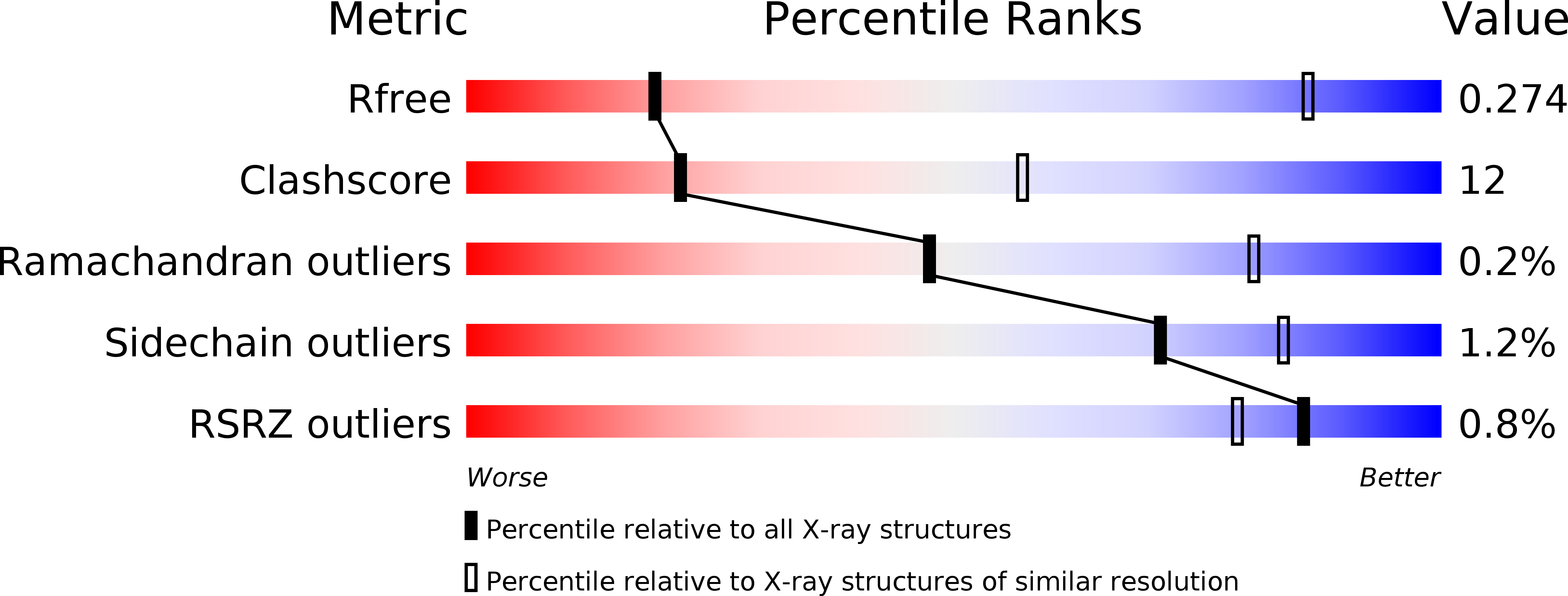
R-Value Free:
0.23
R-Value Work:
0.18
R-Value Observed:
0.19
Space Group:
P 1