
Deposition Date
2017-06-15
Release Date
2017-08-16
Last Version Date
2024-03-13
Entry Detail
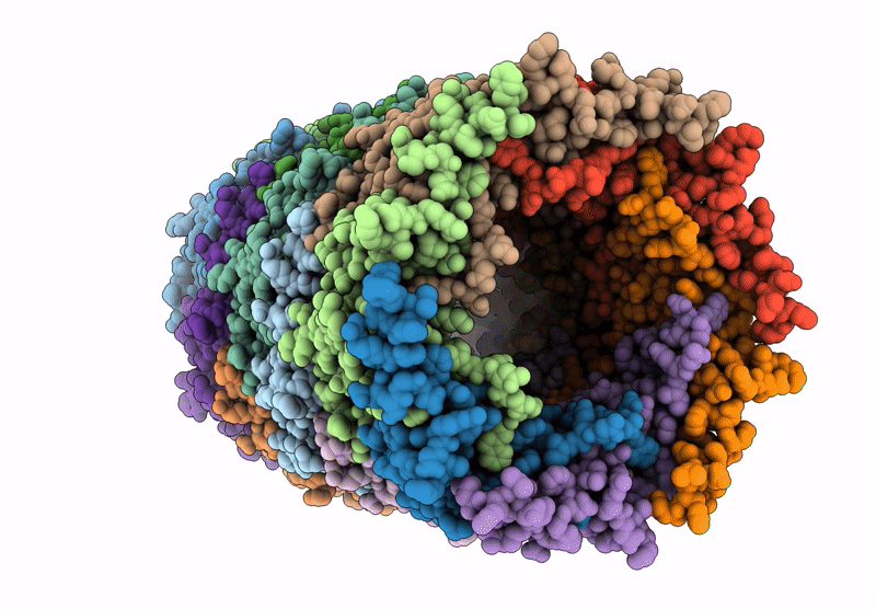
Biological Source:
Source Organism(s):
Enterobacteria phage T4 sensu lato (Taxon ID: 348604)
Method Details:
Experimental Method:
Resolution:
3.40 Å
Aggregation State:
FILAMENT
Reconstruction Method:
HELICAL
