5W4S image
Deposition Date 2017-06-12
Release Date 2018-04-25
Last Version Date 2024-10-30
Entry Detail
PDB ID:
5W4S
Keywords:
Title:
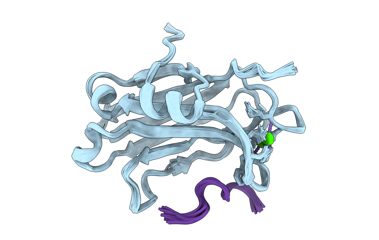
Solution structure of C2 domain from protein kinase C alpha in ternary complex with calcium and V5-pHM peptide
Biological Source:
Source Organism:
Host Organism:
Method Details:
Experimental Method:
Conformers Calculated:
400
Conformers Submitted:
20
Selection Criteria:
structures with the lowest energy
Macromolecular Entities
Polymer Type:polypeptide(L)
Molecule:Protein kinase C alpha type
Gene (Uniprot):Prkca
Chain IDs:A
Chain Length:139
Number of Molecules:1
Biological Source:Rattus norvegicus
Polymer Type:polypeptide(L)
Molecule:V5-pHM peptide
Chain IDs:B
Chain Length:12
Number of Molecules:1
Biological Source:Rattus norvegicus
Primary Citation
Structural Basis of Protein Kinase C alpha Regulation by the C-Terminal Tail.
Biophys. J. 114 1590 1603 (2018)
PMID: 29642029 DOI: 10.1016/j.bpj.2017.12.030

Abstact

Protein kinase C (PKC) isoenzymes are multi-modular proteins activated at the membrane surface to regulate signal transduction processes. When activated by second messengers, PKC undergoes a drastic conformational and spatial transition from the inactive cytosolic state to the activated membrane-bound state. The complete structure of either state of PKC remains elusive. We demonstrate, using NMR spectroscopy, that the isolated Ca2+-sensing membrane-binding C2 domain of the conventional PKCα interacts with a conserved hydrophobic motif of the kinase C-terminal region, and we report a structural model of the complex. Our data suggest that the C-terminal region plays a dual role in regulating the PKC activity: activating, through sensitization of PKC to intracellular Ca2+ oscillations; and auto-inhibitory, through its interaction with a conserved positively charged region of the C2 domain.

Legend

Protein

Chemical

Disease

Primary Citation of related structures