5W3N image
Deposition Date 2017-06-08
Release Date 2017-09-27
Last Version Date 2024-05-15
Entry Detail
PDB ID:
5W3N
Keywords:
Title:
Molecular structure of FUS low sequence complexity domain protein fibrils
Biological Source:
Source Organism:
Homo sapiens (Taxon ID: 9606)
Method Details:
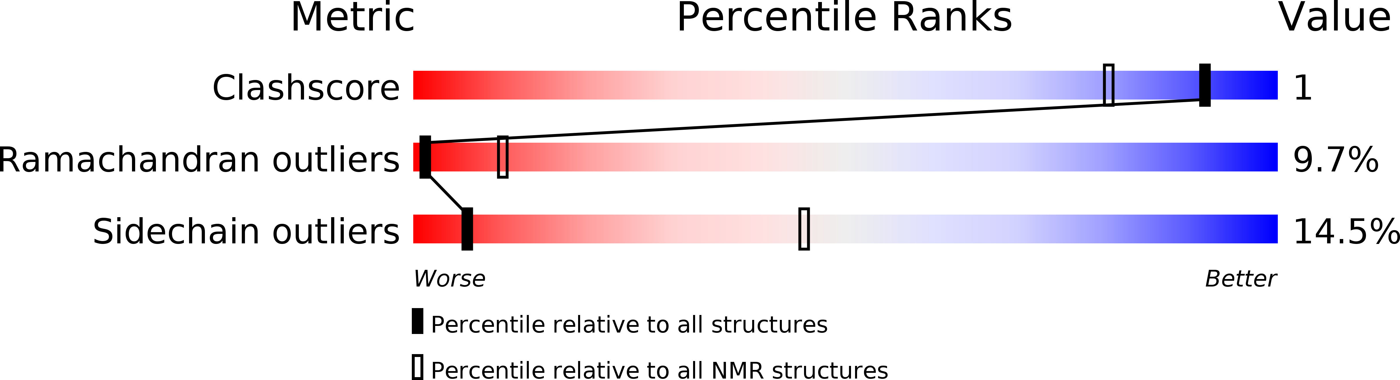
Experimental Method:
Conformers Calculated:
4928
Conformers Submitted:
20
Selection Criteria:
structures with no violations, lowest energy, and derived from one of 44 independent calculations
Macromolecular Entities
Polymer Type:polypeptide(L)
Molecule:RNA-binding protein FUS
Gene (Uniprot):FUS
Chain IDs:A, B, C, D, E, F, G, H, I
Chain Length:241
Number of Molecules:9
Biological Source:Homo sapiens
Ligand Molecules
Primary Citation
Structure of FUS Protein Fibrils and Its Relevance to Self-Assembly and Phase Separation of Low-Complexity Domains.
Cell 171 615 627.e16 (2017)
PMID: 28942918 DOI: 10.1016/j.cell.2017.08.048

Abstact

Polymerization and phase separation of proteins containing low-complexity (LC) domains are important factors in gene expression, mRNA processing and trafficking, and localization of translation. We have used solid-state nuclear magnetic resonance methods to characterize the molecular structure of self-assembling fibrils formed by the LC domain of the fused in sarcoma (FUS) RNA-binding protein. From the 214-residue LC domain of FUS (FUS-LC), a segment of only 57 residues forms the fibril core, while other segments remain dynamically disordered. Unlike pathogenic amyloid fibrils, FUS-LC fibrils lack hydrophobic interactions within the core and are not polymorphic at the molecular structural level. Phosphorylation of core-forming residues by DNA-dependent protein kinase blocks binding of soluble FUS-LC to FUS-LC hydrogels and dissolves phase-separated, liquid-like FUS-LC droplets. These studies offer a structural basis for understanding LC domain self-assembly, phase separation, and regulation by post-translational modification.

Legend

Protein

Chemical

Disease

Primary Citation of related structures