5W0P image
Deposition Date 2017-05-31
Release Date 2017-08-09
Last Version Date 2024-10-30
Entry Detail
PDB ID:
5W0P
Title:
Crystal structure of rhodopsin bound to visual arrestin determined by X-ray free electron laser
Biological Source:
Source Organism:
Host Organism:
Method Details:
Experimental Method:
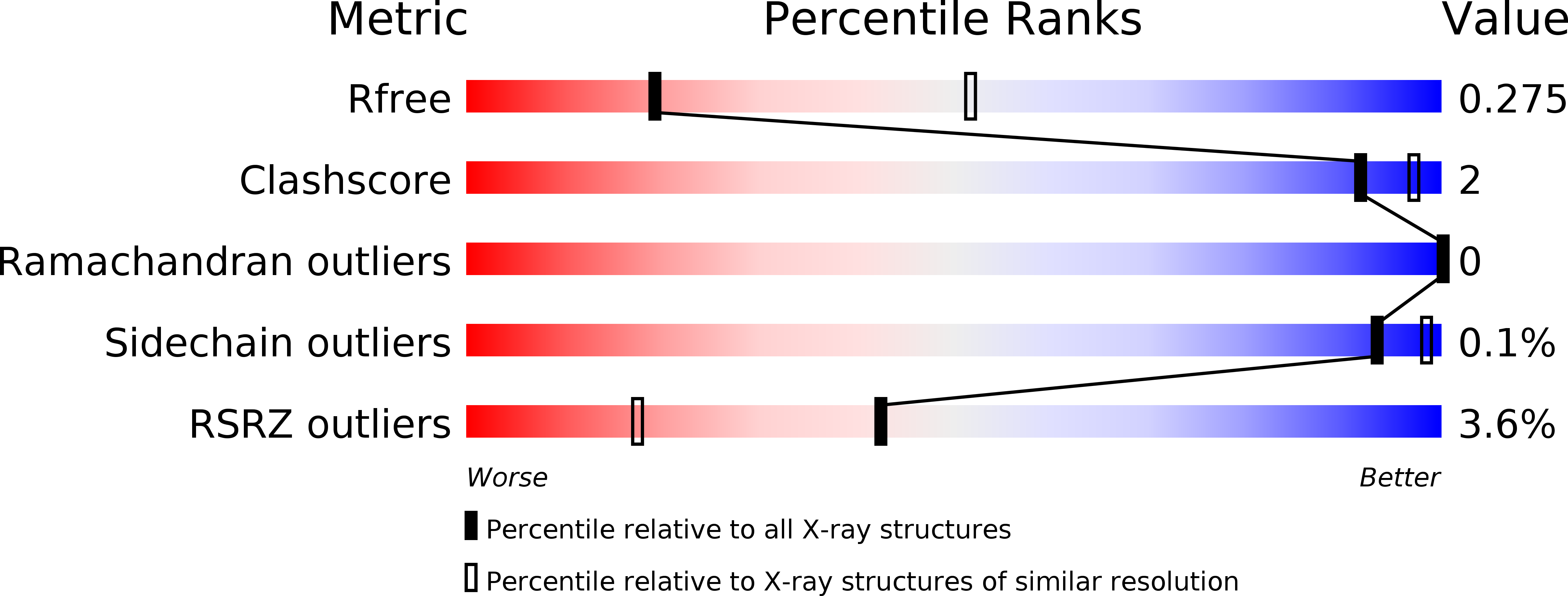
Resolution:
3.01 Å
R-Value Free:
0.27
R-Value Work:
0.23
R-Value Observed:
0.23
Space Group:
P 21 21 21
Macromolecular Entities
Protein Blast
Polymer Type:polypeptide(L)
Molecule:Endolysin,Rhodopsin,S-arrestin
Gene (Uniprot):RHO, Sag
Chain IDs:A, B
Chain Length:906
Number of Molecules:2
Biological Source:Enterobacteria phage RB55, Homo sapiens, Mus musculus
Protein Blast
Polymer Type:polypeptide(L)
Molecule:Endolysin,Rhodopsin,S-arrestin
Gene (Uniprot):RHO, Sag
Chain IDs:C, D
Chain Length:906
Number of Molecules:2
Biological Source:Enterobacteria phage RB55, Homo sapiens, Mus musculus
Modified Residue
Compound ID Chain ID Parent Comp ID Details 2D Image
SEP A SER modified residue
TPO A THR modified residue
Ligand Molecules
Primary Citation
Identification of Phosphorylation Codes for Arrestin Recruitment by G Protein-Coupled Receptors.
Cell 170 457 469.e13 (2017)
PMID: 28753425 DOI: 10.1016/j.cell.2017.07.002

Abstact

G protein-coupled receptors (GPCRs) mediate diverse signaling in part through interaction with arrestins, whose binding promotes receptor internalization and signaling through G protein-independent pathways. High-affinity arrestin binding requires receptor phosphorylation, often at the receptor's C-terminal tail. Here, we report an X-ray free electron laser (XFEL) crystal structure of the rhodopsin-arrestin complex, in which the phosphorylated C terminus of rhodopsin forms an extended intermolecular β sheet with the N-terminal β strands of arrestin. Phosphorylation was detected at rhodopsin C-terminal tail residues T336 and S338. These two phospho-residues, together with E341, form an extensive network of electrostatic interactions with three positively charged pockets in arrestin in a mode that resembles binding of the phosphorylated vasopressin-2 receptor tail to β-arrestin-1. Based on these observations, we derived and validated a set of phosphorylation codes that serve as a common mechanism for phosphorylation-dependent recruitment of arrestins by GPCRs.

Legend

Protein

Chemical

Disease

Primary Citation of related structures