5VYU image
Entry Detail
PDB ID:
5VYU
Keywords:
Title:
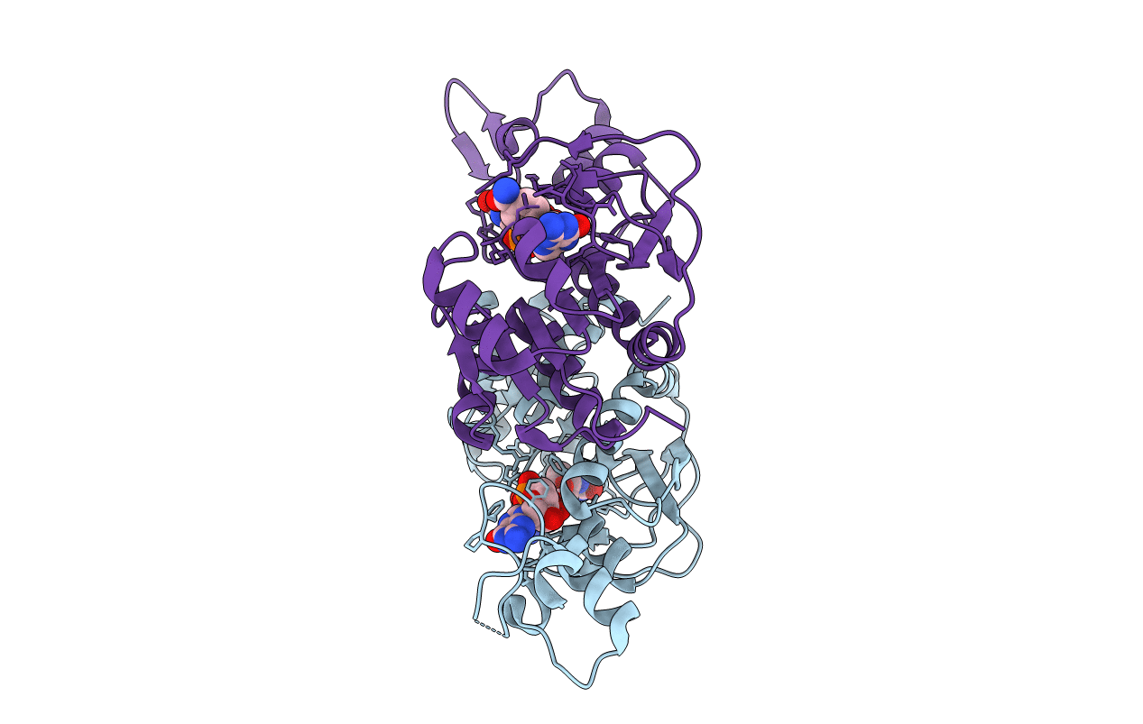
Crystal structure of the WbkC N-formyltransferase from Brucella melitensis in complex with GDP-perosaminea and N-10-formyltetrahydrofolate
Biological Source:
PDB Version:
Deposition Date:
2017-05-26
Release Date:
2017-07-05
Method Details:
Experimental Method:
Resolution:
2.20 Å
R-Value Free:
0.26
R-Value Work:
0.20
R-Value Observed:
0.20
Space Group:
P 43 21 2
Macromolecular Entities
Polymer Type:polypeptide(L)
Description:Gdp-mannose 4,6-dehydratase / gdp-4-amino-4,6-dideoxy-d-mannose formyltransferase
Mutations:D78A, F142A
Chain IDs:A, B
Chain Length:261
Number of Molecules:2
Biological Source:Brucella melitensis biotype 1 (strain 16M / ATCC 23456 / NCTC 10094)
Primary Citation
Biochemical Characterization of WbkC, an N-Formyltransferase from Brucella melitensis.
Biochemistry 56 3657 3668 (2017)
PMID: 28636341 DOI: 10.1021/acs.biochem.7b00494

Abstact

It has become increasingly apparent within the last several years that unusual N-formylated sugars are often found on the O-antigens of such Gram negative pathogenic organisms as Francisella tularensis, Campylobacter jejuni, and Providencia alcalifaciens, among others. Indeed, in some species of Brucella, for example, the O-antigen contains 1,2-linked 4-formamido-4,6-dideoxy-α-d-mannosyl groups. These sugars, often referred to as N-formylperosamine, are synthesized in pathways initiating with GDP-mannose. One of the enzymes required for the production of N-formylperosamine, namely, WbkC, was first identified in 2000 and was suggested to function as an N-formyltransferase. Its biochemical activity was never experimentally verified, however. Here we describe a combined structural and functional investigation of WbkC from Brucella melitensis. Four high resolution X-ray structures of WbkC were determined in various complexes to address its active site architecture. Unexpectedly, the quaternary structure of WbkC was shown to be different from that previously observed for other sugar N-formyltransferases. Additionally, the structures revealed a second binding site for a GDP molecule distinct from that required for GDP-perosamine positioning. In keeping with this additional binding site, kinetic data with the wild type enzyme revealed complex patterns. Removal of GDP binding by mutating Phe 142 to an alanine residue resulted in an enzyme variant displaying normal Michaelis-Menten kinetics. These data suggest that this nucleotide binding pocket plays a role in enzyme regulation. Finally, by using an alternative substrate, we demonstrate that WbkC can be utilized to produce a trideoxysugar not found in nature.

Legend

Protein

Chemical

Disease

Primary Citation of related structures