Deposition Date
2017-05-25
Release Date
2017-08-02
Last Version Date
2024-11-20
Entry Detail
PDB ID:
5VYP
Keywords:
Title:
Crystal structure of the Plant Defensin NsD7 bound to PIP2
Biological Source:
Source Organism(s):
Nicotiana suaveolens (Taxon ID: 200320)
Expression System(s):
Method Details:
Experimental Method:
Resolution:
2.60 Å
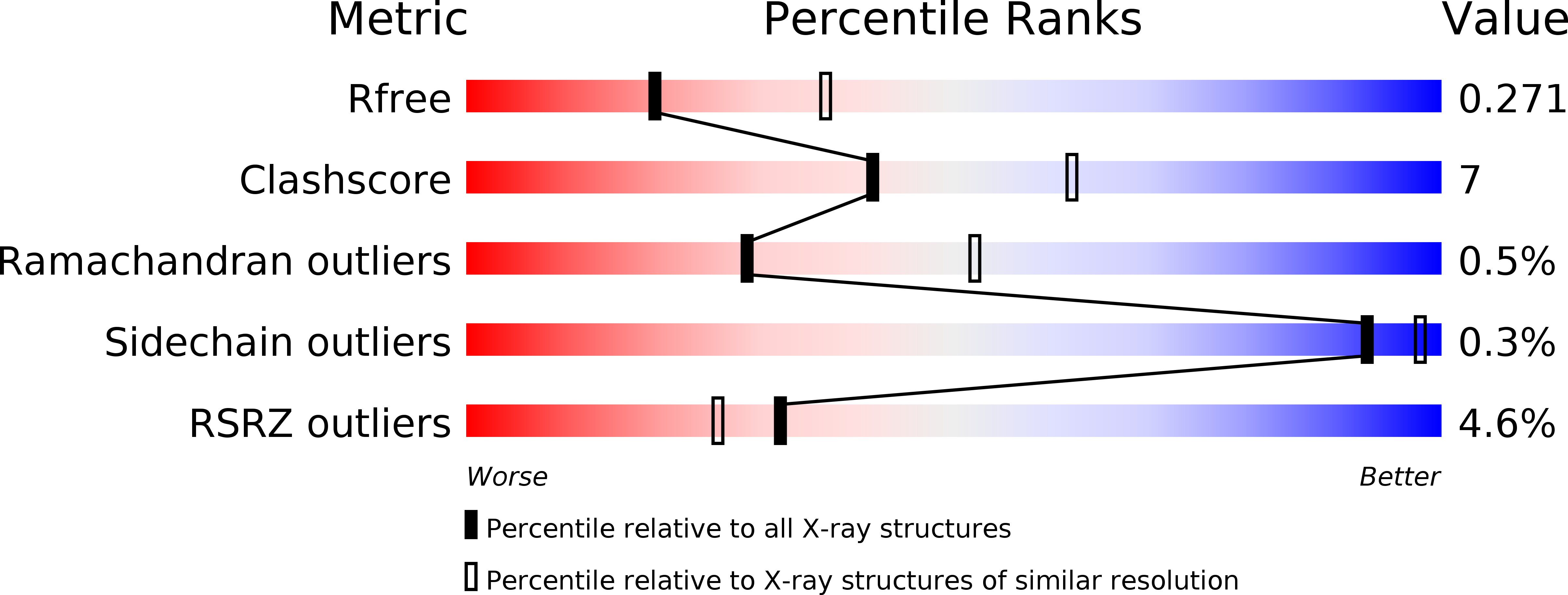
R-Value Free:
0.27
R-Value Work:
0.21
R-Value Observed:
0.21
Space Group:
P 1 21 1