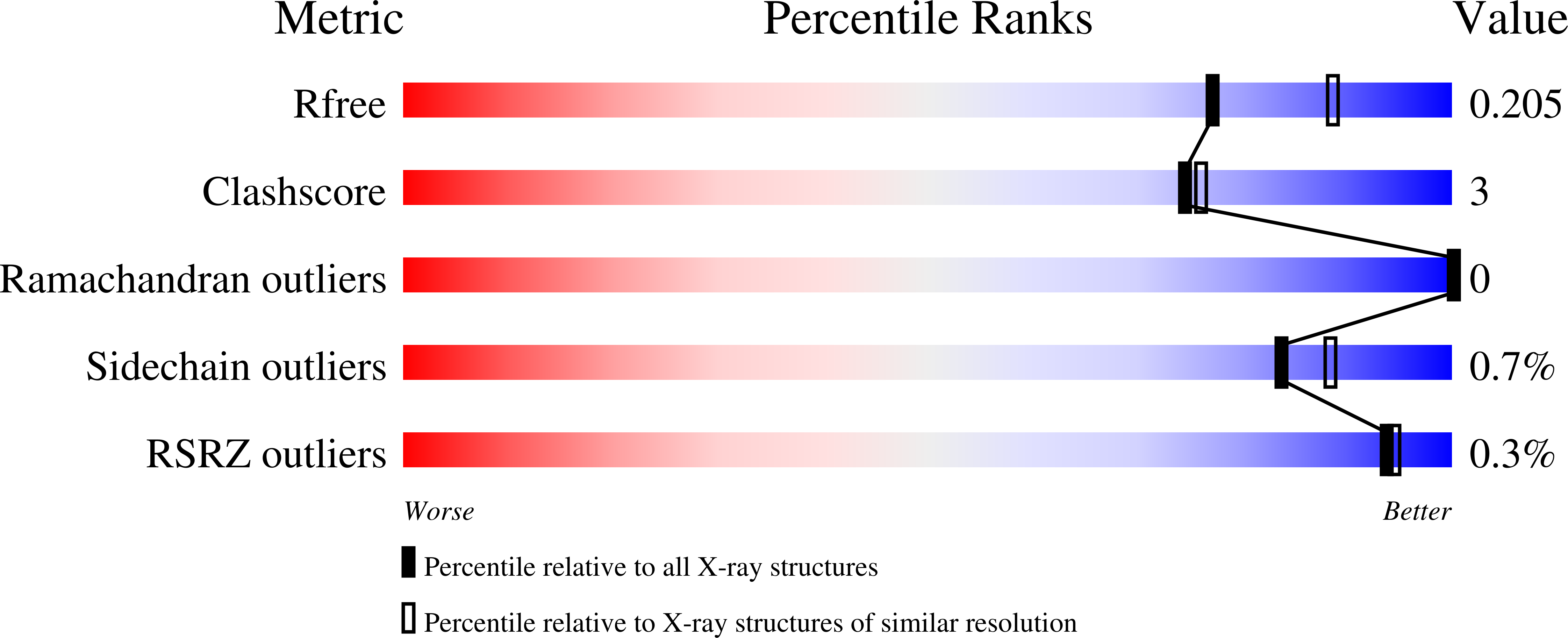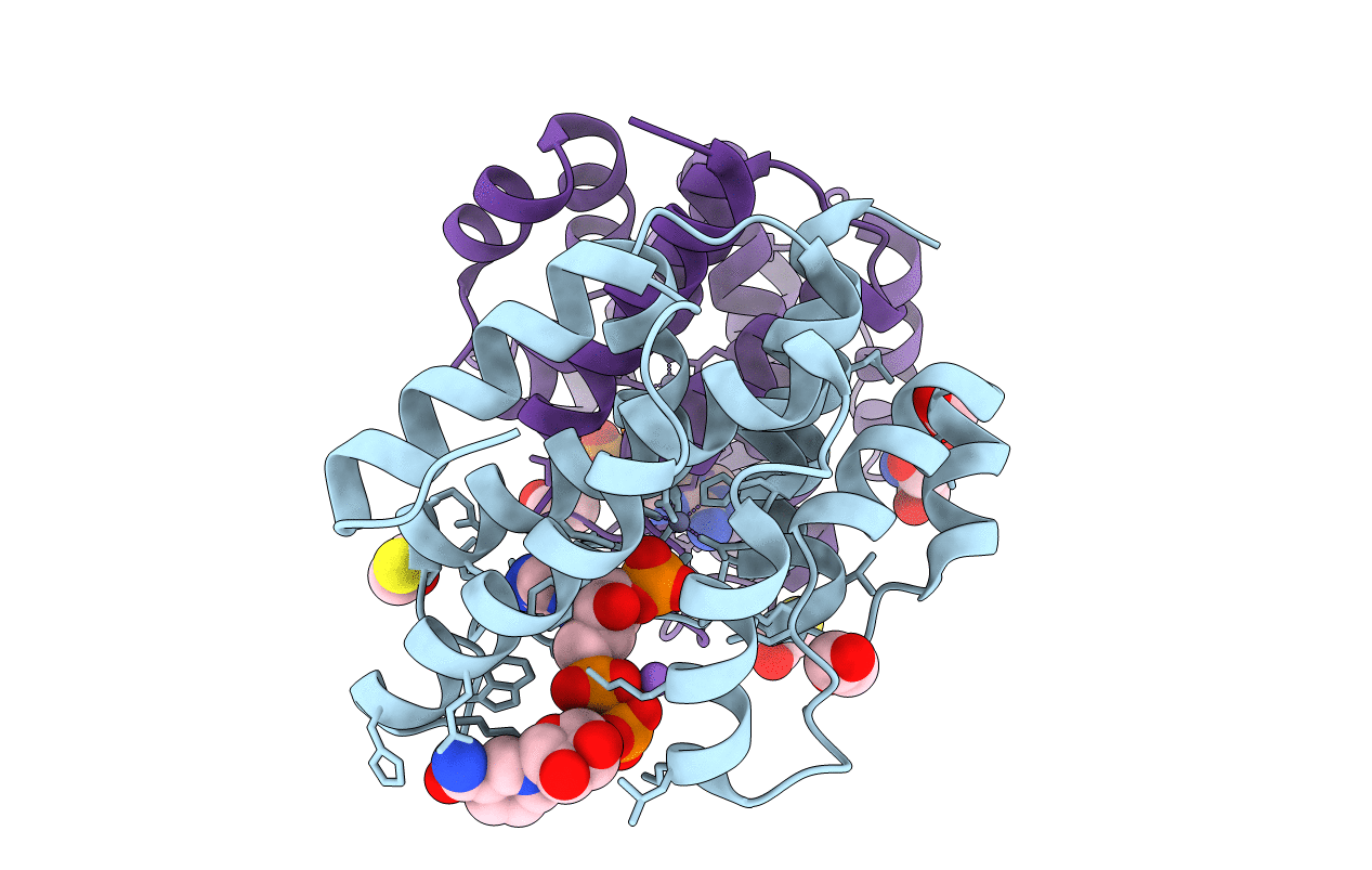
5VXA image
Entry Detail
PDB ID:
5VXA
Keywords:
Title:
Structure of the human Mesh1-NADPH complex
Biological Source:
Source Organism:
Host Organism:
PDB Version:
Deposition Date:
2017-05-23
Release Date:
2018-05-23
Method Details:
Experimental Method:
Resolution:
2.10 Å
R-Value Free:
0.20
R-Value Work:
0.16
R-Value Observed:
0.16
Space Group:
P 21 21 21
Macromolecular Entities
Polymer Type:polypeptide(L)
Description:Guanosine-3',5'-bis(diphosphate) 3'-pyrophosphohydrolase MESH1
Chain IDs:A, B
Chain Length:180
Number of Molecules:2
Biological Source:Homo sapiens
Primary Citation
MESH1 is a cytosolic NADPH phosphatase that regulates ferroptosis.
Nat Metab 2 270 277 (2020)
PMID: 32462112 DOI: 10.1038/s42255-020-0181-1

Abstact

Critical to the bacterial stringent response is the rapid relocation of resources from proliferation toward stress survival through the respective accumulation and degradation of (p)ppGpp by RelA and SpoT homologues. While mammalian genomes encode MESH1, a homologue of the bacterial (p)ppGpp hydrolase SpoT, neither (p)ppGpp nor its synthetase has been identified in mammalian cells. Here, we show that human MESH1 is an efficient cytosolic NADPH phosphatase that facilitates ferroptosis. Visualization of the MESH1-NADPH crystal structure revealed a bona fide affinity for the NADPH substrate. Ferroptosis-inducing erastin or cystine deprivation elevates MESH1, whose overexpression depletes NADPH and sensitizes cells to ferroptosis, whereas MESH1 depletion promotes ferroptosis survival by sustaining the levels of NADPH and GSH and by reducing lipid peroxidation. The ferroptotic protection by MESH1 depletion is ablated by suppression of the cytosolic NAD(H) kinase, NADK, but not its mitochondrial counterpart NADK2. Collectively, these data shed light on the importance of cytosolic NADPH levels and their regulation under ferroptosis-inducing conditions in mammalian cells.

Legend

Protein

Chemical

Disease

Primary Citation of related structures