5VT8 image
Deposition Date 2017-05-15
Release Date 2018-05-23
Last Version Date 2023-10-04
Entry Detail
PDB ID:
5VT8
Keywords:
Title:
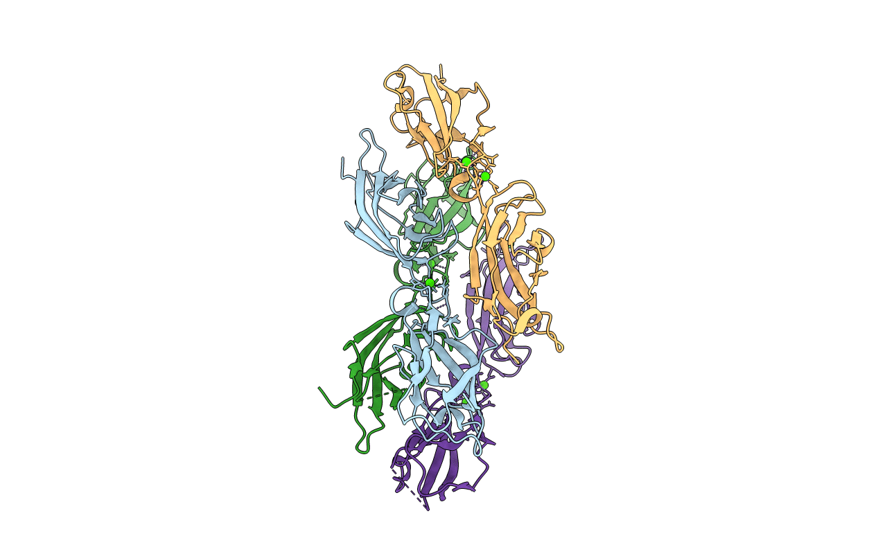
Crystal Structure of Mouse Cadherin-23 EC24-25
Biological Source:
Source Organism:
Mus musculus (Taxon ID: 10090)
Host Organism:
Method Details:
Experimental Method:
Resolution:
2.92 Å
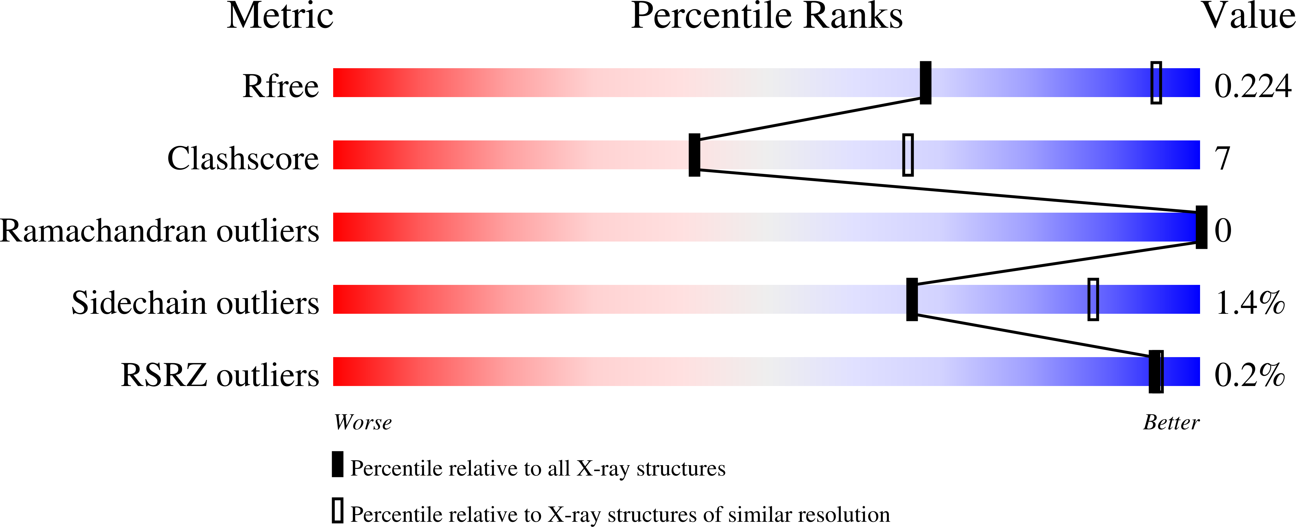
R-Value Free:
0.22
R-Value Work:
0.20
R-Value Observed:
0.20
Space Group:
P 1 21 1
Macromolecular Entities
Structures with similar UniProt ID
Protein Blast
Polymer Type:polypeptide(L)
Molecule:Cadherin-23
Gene (Uniprot):Cdh23
Chain IDs:A (auth: B), B (auth: A), C, D
Chain Length:221
Number of Molecules:4
Biological Source:Mus musculus
Ligand Molecules
Primary Citation
Zooming in on Cadherin-23: Structural Diversity and Potential Mechanisms of Inherited Deafness.
Structure 26 1210 ? (2018)
PMID: 30033219 DOI: 10.1016/j.str.2018.06.003

Abstact

Cadherin-23 (CDH23) is an essential component of hair-cell tip links, fine filaments that mediate inner-ear mechanotransduction. The extracellular domain of CDH23 forms about three-fourths of the tip link with 27 extracellular cadherin (EC) repeats that are structurally similar but not identical to each other. Calcium (Ca2+) coordination at the EC linker regions is key for tip-link elasticity and function. There are ∼116 sites in CDH23 affected by deafness-causing mutations, many of which alter conserved Ca2+-binding residues. Here we present crystal structures showing 18 CDH23 EC repeats, including the most and least conserved, a fragment carrying disease mutations, and EC repeats with non-canonical Ca2+-binding motif sequences and unusual secondary structure. Complementary experiments show deafness mutations' effects on stability and affinity for Ca2+. Additionally, a model of nine contiguous CDH23 EC repeats reveals helicity and potential parallel dimerization faces. Overall, our studies provide detailed structural insight into CDH23 function in mechanotransduction.

Legend

Protein

Chemical

Disease

Primary Citation of related structures