
Deposition Date
2017-05-11
Release Date
2017-06-14
Last Version Date
2024-03-13
Entry Detail
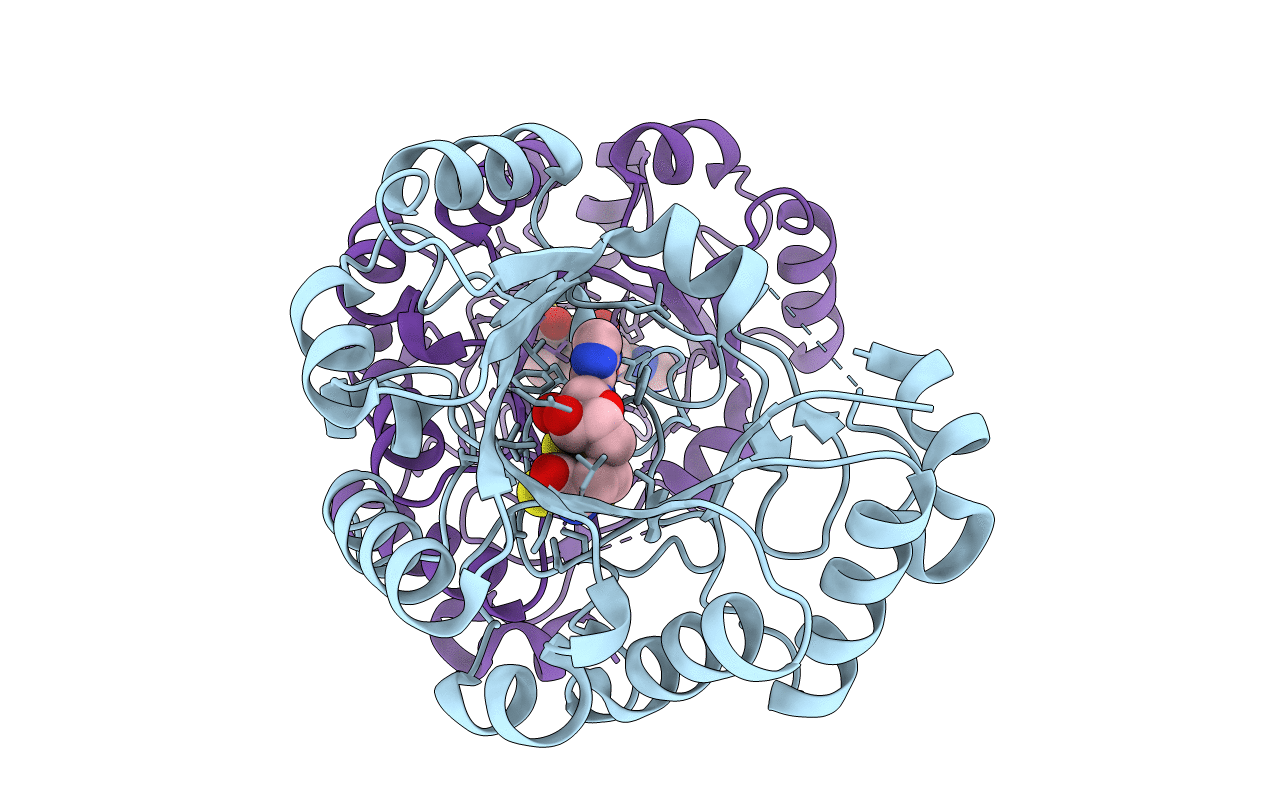
PDB ID:
5VSL
Keywords:
Title:
Crystal structure of viperin with bound [4Fe-4S] cluster and S-adenosylhomocysteine (SAH)
Biological Source:
Source Organism(s):
Mus musculus (Taxon ID: 10090)
Expression System(s):
Method Details:
Experimental Method:
Resolution:
1.97 Å
R-Value Free:
0.19
R-Value Work:
0.15
R-Value Observed:
0.15
Space Group:
P 21 21 21
