5VMM image
Deposition Date 2017-04-27
Release Date 2017-11-15
Last Version Date 2023-10-04
Entry Detail
PDB ID:
5VMM
Title:
Staphylococcus aureus IsdB bound to human hemoglobin
Biological Source:
Source Organism:
Method Details:
Experimental Method:
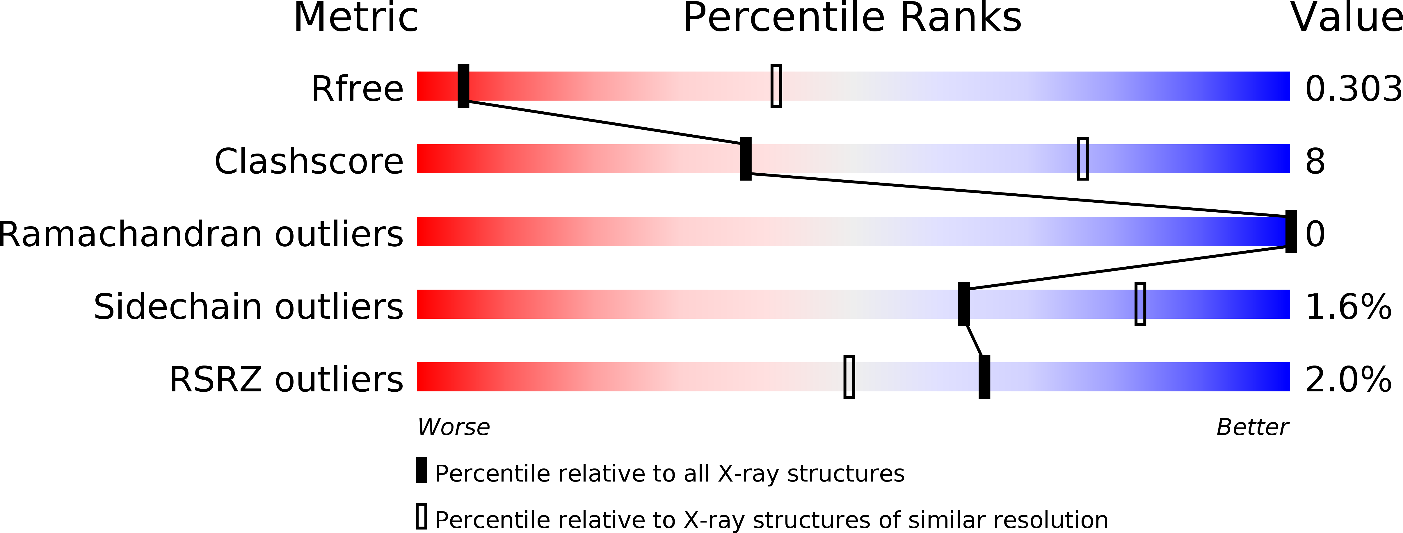
Resolution:
3.60 Å
R-Value Free:
0.30
R-Value Work:
0.25
R-Value Observed:
0.25
Space Group:
P 21 21 2
Macromolecular Entities
Polymer Type:polypeptide(L)
Molecule:Hemoglobin subunit alpha
Gene (Uniprot):HBA1, HBA2
Chain IDs:A, C
Chain Length:141
Number of Molecules:2
Biological Source:Homo sapiens
Polymer Type:polypeptide(L)
Molecule:Hemoglobin subunit beta
Gene (Uniprot):HBB
Chain IDs:B, D
Chain Length:146
Number of Molecules:2
Biological Source:Homo sapiens
Polymer Type:polypeptide(L)
Molecule:Iron-regulated cell wall-anchored protein
Chain IDs:E, F
Chain Length:336
Number of Molecules:2
Biological Source:Staphylococcus aureus
Polymer Type:polypeptide(L)
Molecule:Iron-regulated cell wall-anchored protein
Chain IDs:G (auth: H), H (auth: J)
Chain Length:142
Number of Molecules:2
Biological Source:Staphylococcus aureus
Ligand Molecules
Primary Citation
Structure-function analyses reveal key features in Staphylococcus aureus IsdB-associated unfolding of the heme-binding pocket of human hemoglobin.
J. Biol. Chem. 293 177 190 (2018)
PMID: 29109153 DOI: 10.1074/jbc.M117.806562

Abstact

IsdB is a receptor on the surface of the bacterial pathogen Staphylococcus aureus that extracts heme from hemoglobin (Hb) to enable growth on Hb as a sole iron source. IsdB is critically important both for in vitro growth on Hb and in infection models and is also highly up-regulated in blood, serum, and tissue infection models, indicating a key role of this receptor in bacterial virulence. However, structural information for IsdB is limited. We present here a crystal structure of a complex between human Hb and IsdB. In this complex, the α subunits of Hb are refolded with the heme displaced to the interface with IsdB. We also observe that atypical residues of Hb, His58 and His89 of αHb, coordinate to the heme iron, which is poised for transfer into the heme-binding pocket of IsdB. Moreover, the porphyrin ring interacts with IsdB residues Tyr440 and Tyr444 Previously, Tyr440 was observed to coordinate heme iron in an IsdB·heme complex structure. A Y440F/Y444F IsdB variant we produced was defective in heme transfer yet formed a stable complex with Hb (Kd = 6 ± 2 μm) in solution with spectroscopic features of the bis-His species observed in the crystal structure. Haptoglobin binds to a distinct site on Hb to inhibit heme transfer to IsdB and growth of S. aureus, and a ternary complex of IsdB·Hb·Hp was observed. We propose a model for IsdB heme transfer from Hb that involves unfolding of Hb and heme iron ligand exchange.

Legend

Protein

Chemical

Disease

Primary Citation of related structures