5VJX image
Deposition Date 2017-04-20
Release Date 2017-12-06
Last Version Date 2024-11-13
Entry Detail
PDB ID:
5VJX
Keywords:
Title:
Crystal structure of the CLOCK Transcription Domain Exon19 in Complex with a Repressor
Biological Source:
Source Organism:
Mus musculus (Taxon ID: 10090)
Host Organism:
Method Details:
Experimental Method:
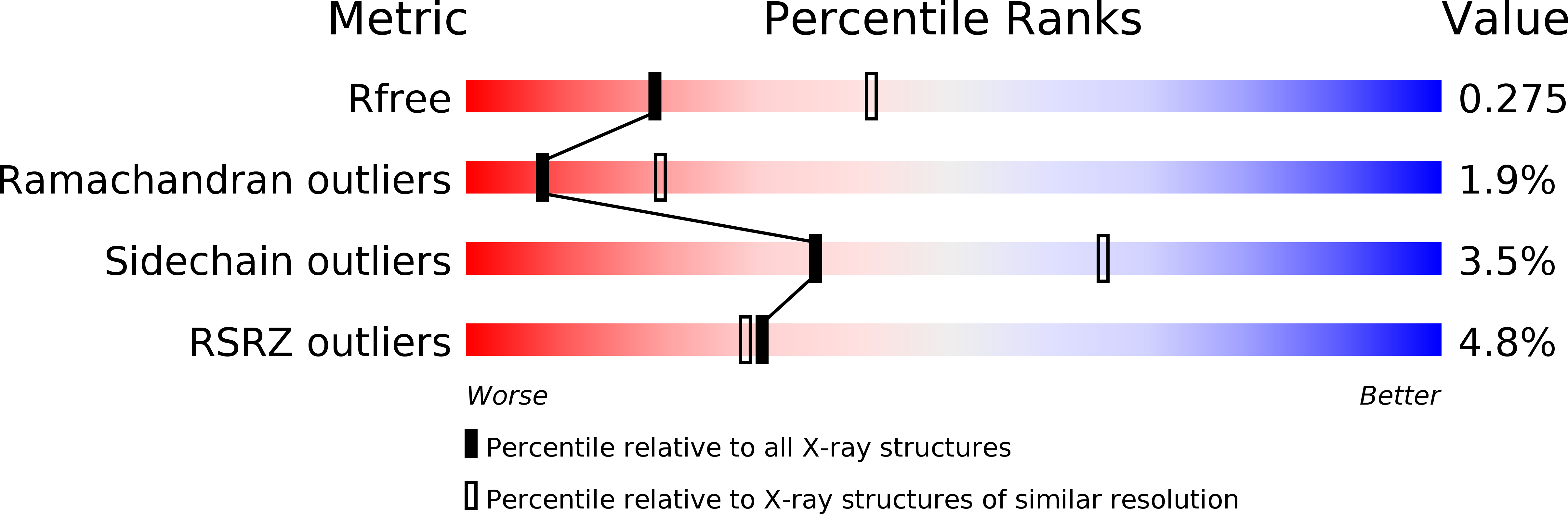
Resolution:
2.70 Å
R-Value Free:
0.27
R-Value Work:
0.21
R-Value Observed:
0.22
Space Group:
C 1 2 1
Macromolecular Entities
Structures with similar UniProt ID
Protein Blast
Polymer Type:polypeptide(L)
Molecule:CLOCK-interacting pacemaker
Gene (Uniprot):Cipc
Chain IDs:A, D, G, J, M, P (auth: R), S (auth: U), V (auth: X), Y (auth: a), BA (auth: d)
Chain Length:64
Number of Molecules:10
Biological Source:Mus musculus
Structures with similar UniProt ID
Protein Blast
Polymer Type:polypeptide(L)
Molecule:Circadian locomoter output cycles protein kaput
Gene (Uniprot):Clock
Chain IDs:B, C, E, F, H, I, K, L, N, O, Q (auth: S), R (auth: T), T (auth: V), U (auth: W), W (auth: Y), X (auth: Z), Z (auth: b), AA (auth: c), CA (auth: e), DA (auth: f)
Chain Length:51
Number of Molecules:20
Biological Source:Mus musculus
Modified Residue
Compound ID Chain ID Parent Comp ID Details 2D Image
MSE A MET modified residue
Primary Citation
Crystal Structure of the CLOCK Transactivation Domain Exon19 in Complex with a Repressor.
Structure 25 1187 1194.e3 (2017)
PMID: 28669630 DOI: 10.1016/j.str.2017.05.023

Abstact

In the canonical clock model, CLOCK:BMAL1-mediated transcriptional activation is feedback regulated by its repressors CRY and PER and, in association with other coregulators, ultimately generates oscillatory gene expression patterns. How CLOCK:BMAL1 interacts with coregulator(s) is not well understood. Here we report the crystal structures of the mouse CLOCK transactivating domain Exon19 in complex with CIPC, a potent circadian repressor that functions independently of CRY and PER. The Exon19:CIPC complex adopts a three-helical coiled-coil bundle conformation containing two Exon19 helices and one CIPC. Unique to Exon19:CIPC, three highly conserved polar residues, Asn341 of CIPC and Gln544 of the two Exon19 helices, are located at the mid-section of the coiled-coil bundle interior and form hydrogen bonds with each other. Combining results from protein database search, sequence analysis, and mutagenesis studies, we discovered for the first time that CLOCK Exon19:CIPC interaction is a conserved transcription regulatory mechanism among mammals, fish, flies, and other invertebrates.

Legend

Protein

Chemical

Disease

Primary Citation of related structures