5VIV image
Entry Detail
PDB ID:
5VIV
Title:
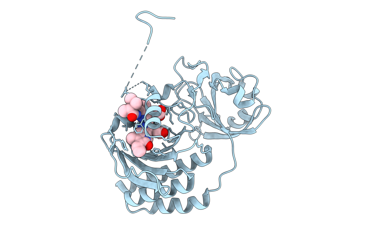
Crystal structure of monomeric near-infrared fluorescent protein miRFP670
Biological Source:
Host Organism:
PDB Version:
Deposition Date:
2017-04-17
Release Date:
2017-06-21
Method Details:
Experimental Method:
Resolution:
1.33 Å
R-Value Free:
0.18
R-Value Work:
0.15
R-Value Observed:
0.15
Space Group:
P 21 21 21
Macromolecular Entities
Polymer Type:polypeptide(L)
Description:monomeric near-infrared fluorescent protein miRFP670
Chain IDs:A
Chain Length:315
Number of Molecules:1
Biological Source:Rhodopseudomonas palustris
Primary Citation
Designing brighter near-infrared fluorescent proteins: insights from structural and biochemical studies.
Chem Sci 8 4546 4557 (2017)
PMID: 28936332 DOI: 10.1039/c7sc00855d

Abstact

Brighter near-infrared (NIR) fluorescent proteins (FPs) are required for multicolor microscopy and deep-tissue imaging. Here, we present structural and biochemical analyses of three monomeric, spectrally distinct phytochrome-based NIR FPs, termed miRFPs. The miRFPs are closely related and differ by only a few amino acids, which define their molecular brightness, brightness in mammalian cells, and spectral properties. We have identified the residues responsible for the spectral red-shift, revealed a new chromophore bound simultaneously to two cysteine residues in the PAS and GAF domains in blue-shifted NIR FPs, and uncovered the importance of amino acid residues in the N-terminus of NIR FPs for their molecular and cellular brightness. The novel chromophore covalently links the N-terminus of NIR FPs with their C-terminal GAF domain, forming a topologically closed knot in the structure, and also contributes to the increased brightness. Based on our studies, we suggest a strategy to develop spectrally distinct NIR FPs with enhanced brightness.

Legend

Protein

Chemical

Disease

Primary Citation of related structures