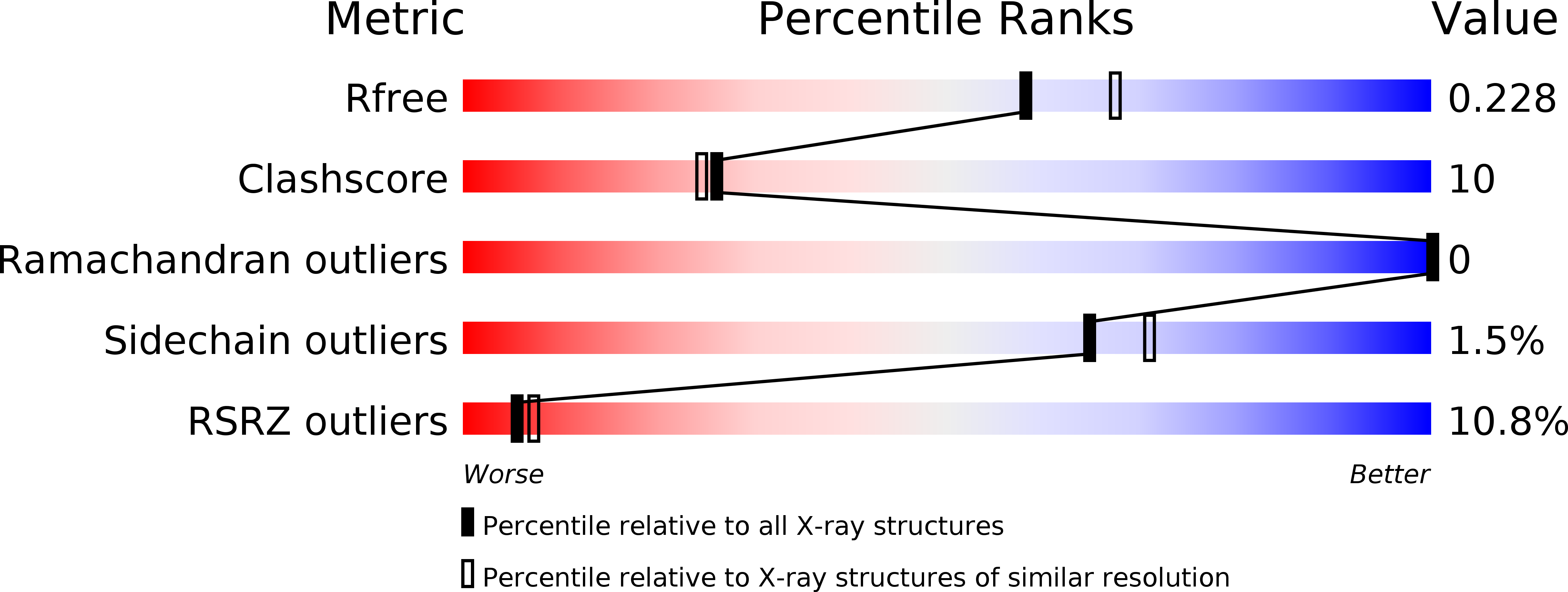
5VGR image
Deposition Date 2017-04-11
Release Date 2017-05-17
Last Version Date 2023-10-04
Entry Detail
PDB ID:
5VGR
Keywords:
Title:
Human Atlastin-3, GDP-bound
Biological Source:
Source Organism:
Homo sapiens (Taxon ID: 9606)
Host Organism:
Method Details:
Experimental Method:
Resolution:
2.10 Å
R-Value Free:
0.22
R-Value Work:
0.19
R-Value Observed:
0.19
Space Group:
C 1 2 1
Macromolecular Entities
Polymer Type:polypeptide(L)
Molecule:Atlastin-3
Gene (Uniprot):ATL3
Chain IDs:A, B
Chain Length:431
Number of Molecules:2
Biological Source:Homo sapiens
Ligand Molecules
Primary Citation
Timing and Reset Mechanism of GTP Hydrolysis-Driven Conformational Changes of Atlastin.
Structure 25 997 1010.e4 (2017)
PMID: 28602821 DOI: 10.1016/j.str.2017.05.007

Abstact

The endoplasmic reticulum (ER) forms a branched, dynamic membrane tubule network that is vital for cellular function. Branching arises from membrane fusion facilitated by the GTPase atlastin (ATL). Many metazoan genomes encode for three ATL isoforms that appear to fulfill partially redundant function despite differences in their intrinsic GTPase activity and localization within the ER; however, the underlying mechanistic differences between the isoforms are poorly understood. Here, we identify discrete temporal steps in the catalytic cycle for the two most dissimilar isoforms, ATL1 and ATL3, revealing an overall conserved progression of molecular events from nucleotide binding and hydrolysis to ATL dimerization and phosphate release. A crystal structure of ATL3 suggests a mechanism for the displacement of the catalytic Mg2+ ion following guanosine triphosphate (GTP) hydrolysis. Together, the data extend the mechanistic framework for how GTP hydrolysis drives conformational changes in ATL and how the cycle is reset for subsequent rounds of catalysis.

Legend

Protein

Chemical

Disease

Primary Citation of related structures