
Deposition Date
2017-04-10
Release Date
2017-05-24
Last Version Date
2024-11-13
Entry Detail
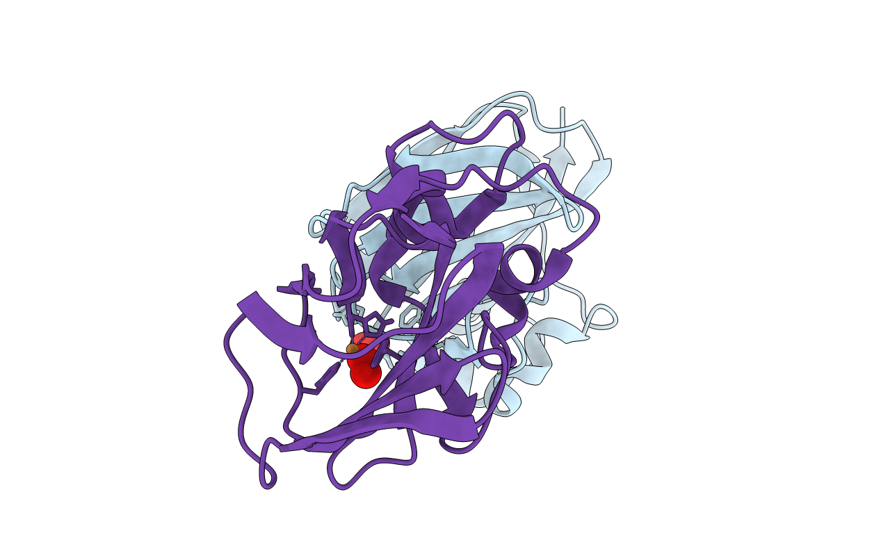
PDB ID:
5VG0
Keywords:
Title:
Room temperature X-ray crystallographic structure of a Jonesia denitrificans lytic polysaccharide monooxygenase at 1.1 angstrom resolution.
Biological Source:
Source Organism(s):
Expression System(s):
Method Details:
Experimental Method:
Resolution:
1.10 Å
R-Value Free:
0.12
R-Value Work:
0.11
R-Value Observed:
0.11
Space Group:
P 21 21 21
