5VFF image
Deposition Date 2017-04-07
Release Date 2018-04-11
Last Version Date 2023-10-04
Entry Detail
PDB ID:
5VFF
Title:
Synaptotagmin 1 C2B domain, lead-bound (low occupancy)
Biological Source:
Source Organism:
Mus musculus (Taxon ID: 10090)
Host Organism:
Method Details:
Experimental Method:
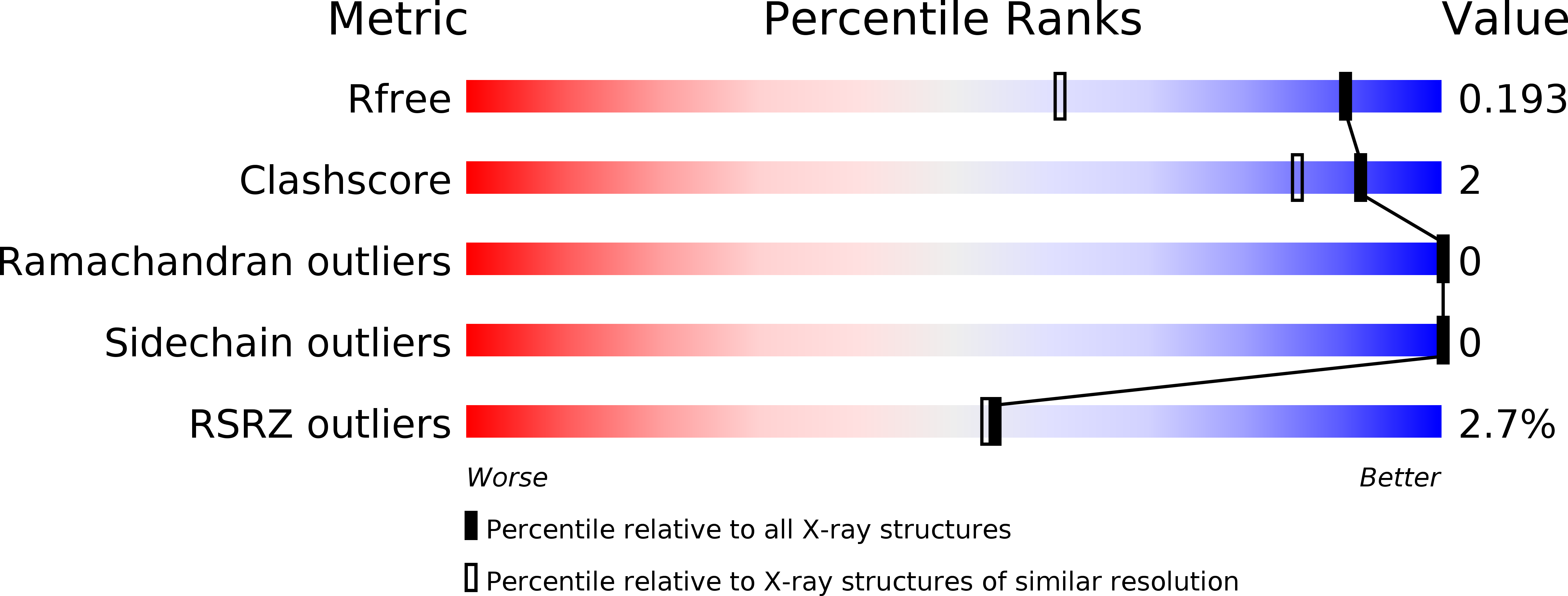
Resolution:
1.41 Å
R-Value Free:
0.19
R-Value Work:
0.15
R-Value Observed:
0.15
Space Group:
P 21 21 21
Macromolecular Entities
Polymer Type:polypeptide(L)
Molecule:Synaptotagmin-1
Gene (Uniprot):Syt1
Mutations:C277S
Chain IDs:A
Chain Length:151
Number of Molecules:1
Biological Source:Mus musculus
Primary Citation
High affinity interactions of Pb2+with synaptotagmin I.
Metallomics 10 1211 1222 (2018)
PMID: 30063057 DOI: 10.1039/c8mt00135a

Abstact

Lead (Pb) is a potent neurotoxin that disrupts synaptic neurotransmission. We report that Synaptotagmin I (SytI), a key regulator of Ca2+-evoked neurotransmitter release, has two high-affinity Pb2+ binding sites that belong to its cytosolic C2A and C2B domains. The crystal structures of Pb2+-complexed C2 domains revealed that protein-bound Pb2+ ions have holodirected coordination geometries and all-oxygen coordination spheres. The on-rate constants of Pb2+ binding to the C2 domains of SytI are comparable to those of Ca2+ and are diffusion-limited. In contrast, the off-rate constants are at least two orders of magnitude smaller, indicating that Pb2+ can serve as both a thermodynamic and kinetic trap for the C2 domains. We demonstrate, using NMR spectroscopy, that population of these sites by Pb2+ ions inhibits further Ca2+ binding despite the existing coordination vacancies. Our work offers a unique insight into the bioinorganic chemistry of Pb(ii) and suggests a mechanism by which low concentrations of Pb2+ ions can interfere with the Ca2+-dependent function of SytI in the cell.

Legend

Protein

Chemical

Disease

Primary Citation of related structures