Deposition Date
2017-04-04
Release Date
2018-06-27
Last Version Date
2023-10-04
Entry Detail
PDB ID:
5VEC
Keywords:
Title:
Crystal Structure of the R515L missense variant of human PGM1
Biological Source:
Source Organism(s):
Homo sapiens (Taxon ID: 9606)
Expression System(s):
Method Details:
Experimental Method:
Resolution:
2.20 Å
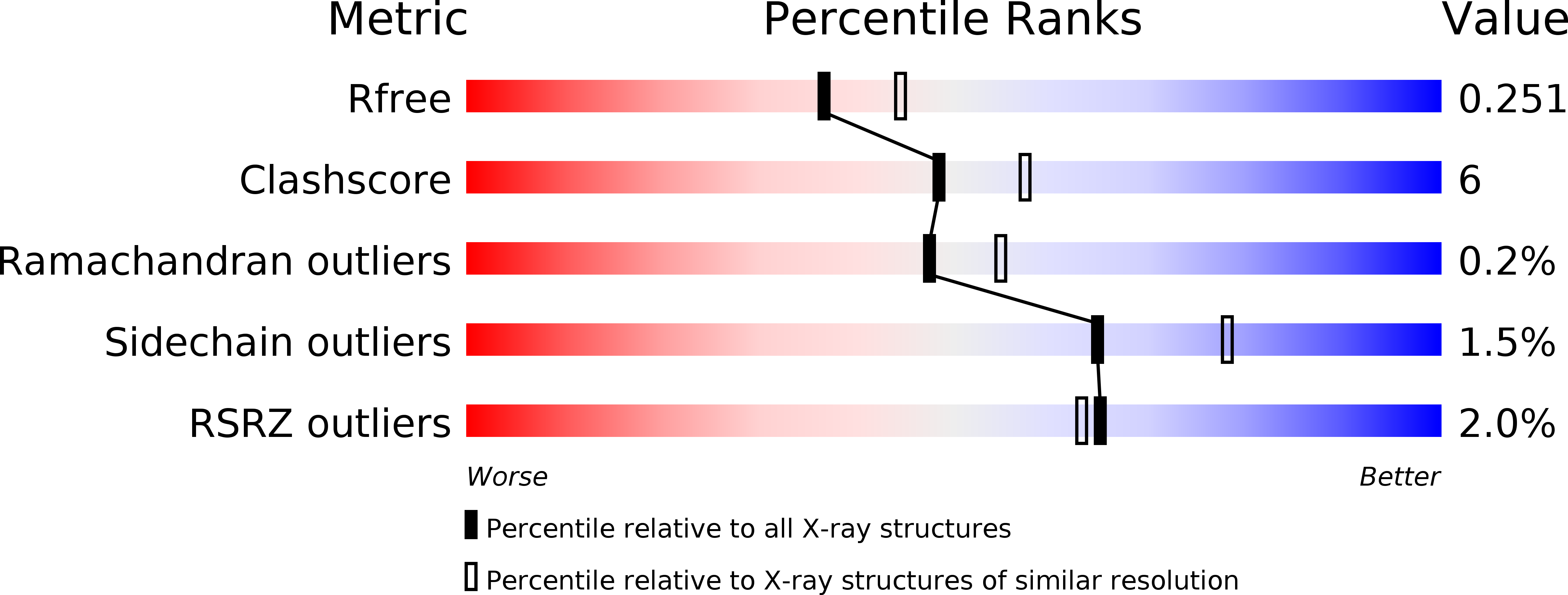
R-Value Free:
0.25
R-Value Work:
0.18
R-Value Observed:
0.19
Space Group:
P 41 21 2