5VDH image
Deposition Date 2017-04-03
Release Date 2017-05-24
Last Version Date 2024-10-23
Entry Detail
PDB ID:
5VDH
Title:
Crystal Structure of Human Glycine Receptor alpha-3 Bound to AM-3607, Glycine, and Ivermectin
Biological Source:
Source Organism:
Homo sapiens (Taxon ID: 9606)
Host Organism:
Method Details:
Experimental Method:
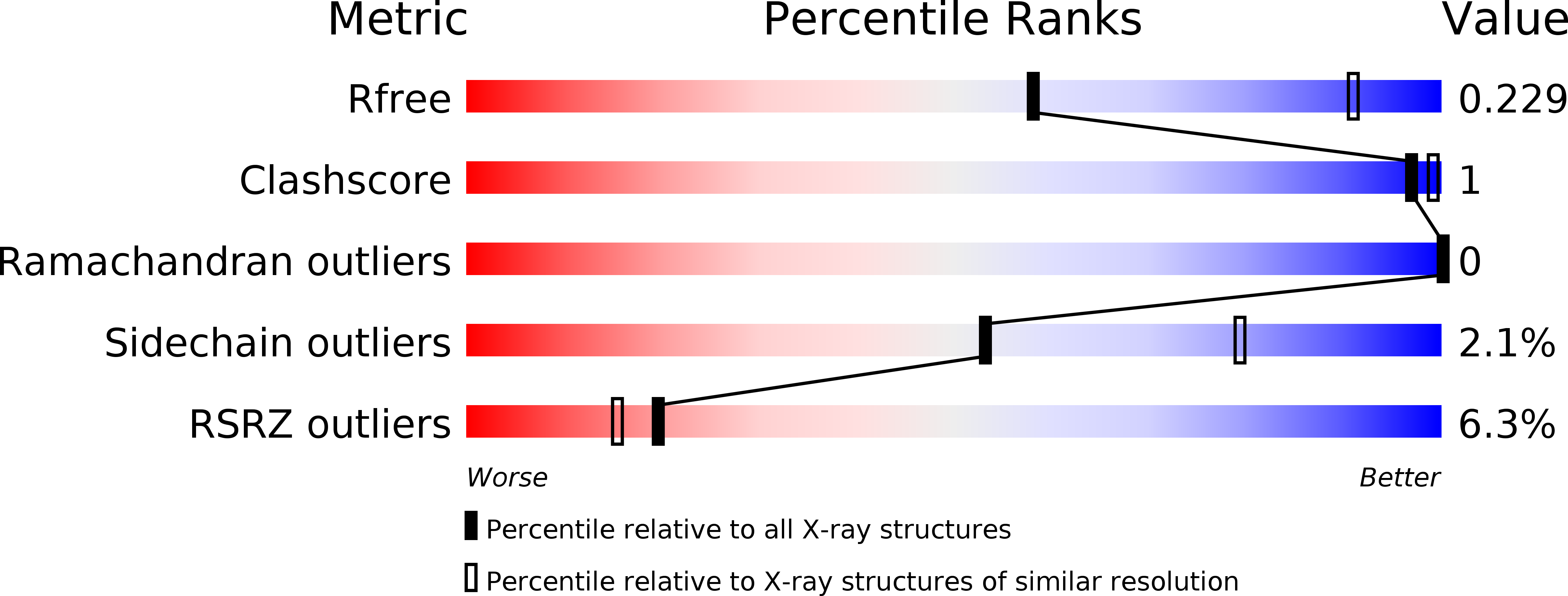
Resolution:
2.85 Å
R-Value Free:
0.23
R-Value Work:
0.22
R-Value Observed:
0.22
Space Group:
P 43 21 2
Macromolecular Entities
Structures with similar UniProt ID
Protein Blast
Polymer Type:polypeptide(L)
Molecule:Glycine receptor subunit alpha-3
Gene (Uniprot):GLRA3
Chain IDs:A, B, C, D, E
Chain Length:362
Number of Molecules:5
Biological Source:Homo sapiens
Primary Citation
Crystal Structures of Human GlyR alpha 3 Bound to Ivermectin.
Structure 25 945 950.e2 (2017)
PMID: 28479061 DOI: 10.1016/j.str.2017.04.007

Abstact

Ivermectin acts as a positive allosteric modulator of several Cys-loop receptors including the glutamate-gated chloride channels (GluCls), γ-aminobutyric acid receptors (GABAARs), glycine receptors (GlyRs), and neuronal α7-nicotinic receptors (α7 nAChRs). The crystal structure of Caenorhabditis elegans GluCl complexed with ivermectin revealed the details of its ivermectin binding site. Although the electron microscopy structure of zebrafish GlyRα1 complexed with ivermectin demonstrated a similar binding orientation, detailed structural information on the ivermectin binding and pore opening for Cys-loop receptors in vertebrates has been elusive. Here we present the crystal structures of human GlyRα3 in complex with ivermectin at 2.85 and 3.08 Å resolution. Our structures allow us to explore in detail the molecular recognition of ivermectin by GlyRs, GABAARs, and α7 nAChRs. Comparisons with previous structures reveal how the ivermectin binding expands the ion channel pore. Our results hold promise in structure-based design of GlyR modulators for the treatment of neuropathic pain.

Legend

Protein

Chemical

Disease

Primary Citation of related structures