5VBS image
Deposition Date 2017-03-30
Release Date 2018-03-07
Last Version Date 2023-10-04
Entry Detail
PDB ID:
5VBS
Keywords:
Title:
Structural basis for a six letter alphabet including GATCKX
Biological Source:
Method Details:
Experimental Method:
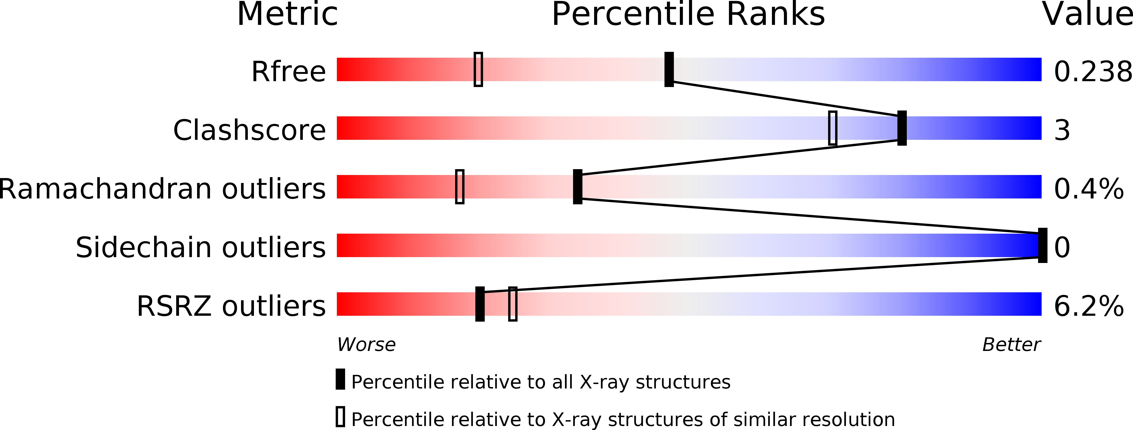
Resolution:
1.75 Å
R-Value Free:
0.23
R-Value Work:
0.20
R-Value Observed:
0.21
Space Group:
P 21 21 2
Macromolecular Entities
Structures with similar UniProt ID
Protein Blast
Polymer Type:polypeptide(L)
Molecule:reverse transcriptase catalytic fragment
Gene (Uniprot):gag-pol
Chain IDs:A
Chain Length:259
Number of Molecules:1
Biological Source:Moloney murine leukemia virus (isolate Shinnick)
Polymer Type:polydeoxyribonucleotide
Molecule:DNA (5'-D(*CP*TP*TP*AP*TP*(DX)P*(DX)P*T)-3')
Chain IDs:B
Chain Length:8
Number of Molecules:1
Biological Source:Escherichia coli
Polymer Type:polydeoxyribonucleotide
Molecule:DNA (5'-D(P*AP*(93D)P*(93D)P*AP*TP*AP*AP*G)-3')
Chain IDs:C (auth: G)
Chain Length:8
Number of Molecules:1
Biological Source:Escherichia coli
Primary Citation
Structure and Biophysics for a Six Letter DNA Alphabet that Includes Imidazo[1,2-a]-1,3,5-triazine-2(8H)-4(3H)-dione (X) and 2,4-Diaminopyrimidine (K).
ACS Synth Biol 6 2118 2129 (2017)
PMID: 28752992 DOI: 10.1021/acssynbio.7b00150

Abstact

A goal of synthetic biology is to develop new nucleobases that retain the desirable properties of natural nucleobases at the same time as expanding the genetic alphabet. The nonstandard Watson-Crick pair between imidazo[1,2-a]-1,3,5-triazine-2(8H)-4(3H)-dione (X) and 2,4-diaminopyrimidine (K) does exactly this, pairing via complementary arrangements of hydrogen bonding in these two nucleobases, which do not complement any natural nucleobase. Here, we report the crystal structure of a duplex DNA oligonucleotide in B-form including two consecutive X:K pairs in GATCXK DNA determined as a host-guest complex at 1.75 Å resolution. X:K pairs have significant propeller twist angles, similar to those observed for A:T pairs, and a calculated hydrogen bonding pairing energy that is weaker than that of A:T. Thus, although inclusion of X:K pairs results in a duplex DNA structure that is globally similar to that of an analogous G:C structure, the X:K pairs locally and energetically more closely resemble A:T pairs.

Legend

Protein

Chemical

Disease

Primary Citation of related structures