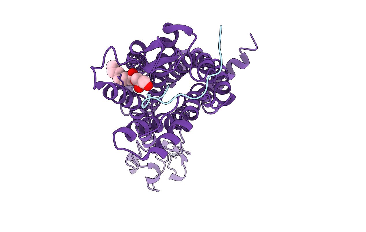
5VBL image
Entry Detail
PDB ID:
5VBL
Title:
Structure of apelin receptor in complex with agonist peptide
Biological Source:
PDB Version:
Deposition Date:
2017-03-29
Release Date:
2017-05-31
Method Details:
Experimental Method:
Resolution:
2.60 Å
R-Value Free:
0.25
R-Value Work:
0.23
R-Value Observed:
0.24
Space Group:
C 1 2 1
Macromolecular Entities
Polymer Type:polypeptide(L)
Description:agonist peptide
Chain IDs:A
Chain Length:17
Number of Molecules:1
Biological Source:Homo sapiens
Polymer Type:polypeptide(L)
Description:Apelin receptor,Rubredoxin,Apelin receptor Chimera
Mutations:V117A, T177N,W261K, C325L, C326M
Chain IDs:B
Chain Length:407
Number of Molecules:1
Biological Source:Homo sapiens, Clostridium pasteurianum
Primary Citation
Structural Basis for Apelin Control of the Human Apelin Receptor
Structure 25 858 866.e4 (2017)
PMID: 28528775 DOI: 10.1016/j.str.2017.04.008

Abstact

Apelin receptor (APJR) is a key regulator of human cardiovascular function and is activated by two different endogenous peptide ligands, apelin and Elabela, each with different isoforms diversified by length and amino acid sequence. Here we report the 2.6-Å resolution crystal structure of human APJR in complex with a designed 17-amino-acid apelin mimetic peptide agonist. The structure reveals that the peptide agonist adopts a lactam constrained curved two-site ligand binding mode. Combined with mutation analysis and molecular dynamics simulations with apelin-13 binding to the wild-type APJR, this structure provides a mechanistic understanding of apelin recognition and binding specificity. Comparison of this structure with that of other peptide receptors suggests that endogenous peptide ligands with a high degree of conformational flexibility may bind and modulate the receptors via a similar two-site binding mechanism.

Legend

Protein

Chemical

Disease

Primary Citation of related structures