5V6J image
Deposition Date 2017-03-16
Release Date 2017-08-16
Last Version Date 2024-10-23
Entry Detail
PDB ID:
5V6J
Title:
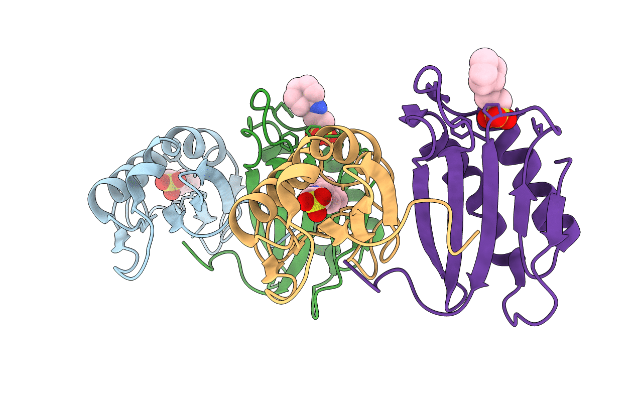
Glycan binding protein Y3 from mushroom Coprinus comatus possesses anti-leukemic activity
Biological Source:
Source Organism:
Host Organism:
Method Details:
Experimental Method:
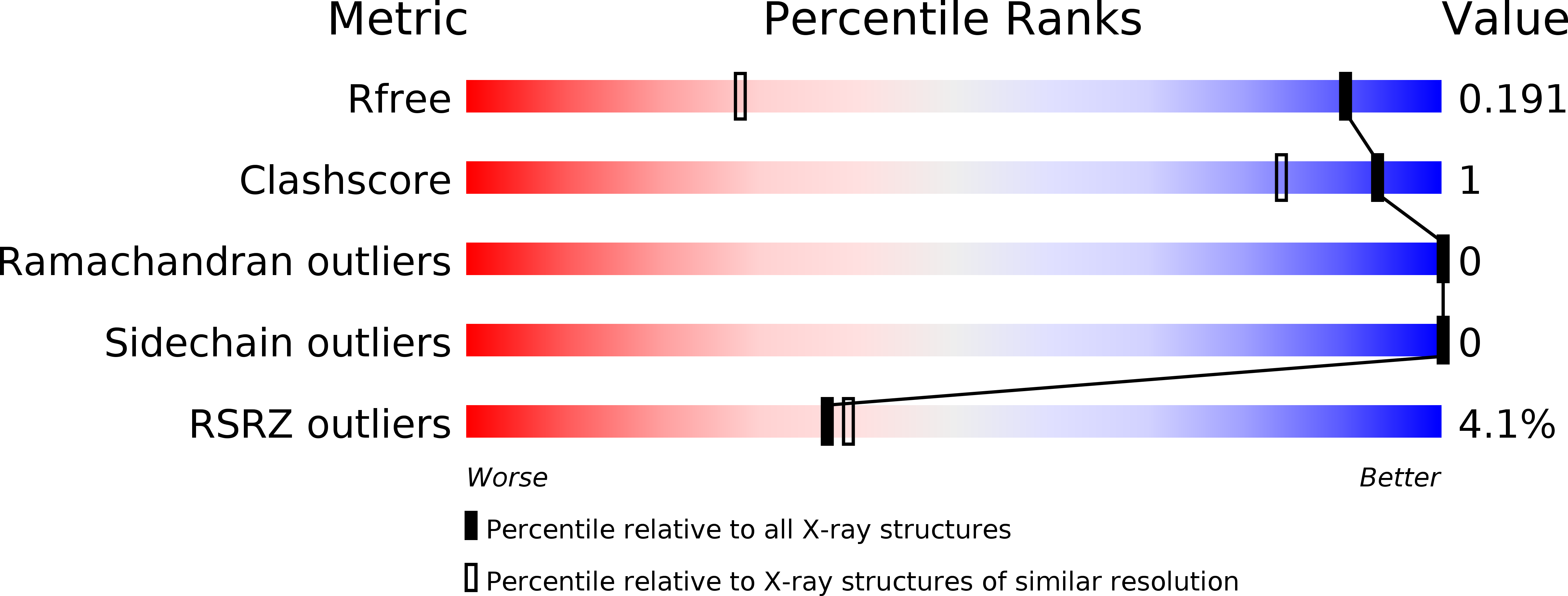
Resolution:
1.18 Å
R-Value Free:
0.18
R-Value Work:
0.16
Space Group:
P 1 21 1
Macromolecular Entities
Structures with similar UniProt ID
Protein Blast
Polymer Type:polypeptide(L)
Molecule:TMV resistance protein Y3
Gene (Uniprot):y3
Chain IDs:A, B, C, D
Chain Length:112
Number of Molecules:4
Biological Source:Coprinus comatus
Modified Residue
Compound ID Chain ID Parent Comp ID Details 2D Image
PCA A GLN modified residue
Ligand Molecules
Primary Citation
Cytotoxic protein from the mushroom Coprinus comatus possesses a unique mode for glycan binding and specificity.
Proc. Natl. Acad. Sci. U.S.A. 114 8980 8985 (2017)
PMID: 28784797 DOI: 10.1073/pnas.1706894114

Abstact

Glycans possess significant chemical diversity; glycan binding proteins (GBPs) recognize specific glycans to translate their structures to functions in various physiological and pathological processes. Therefore, the discovery and characterization of novel GBPs and characterization of glycan-GBP interactions are significant to provide potential targets for therapeutic intervention of many diseases. Here, we report the biochemical, functional, and structural characterization of a 130-amino-acid protein, Y3, from the mushroom Coprinus comatus Biochemical studies of recombinant Y3 from a yeast expression system demonstrated the protein is a unique GBP. Additionally, we show that Y3 exhibits selective and potent cytotoxicity toward human T-cell leukemia Jurkat cells compared with a panel of cancer cell lines via inducing caspase-dependent apoptosis. Screening of a glycan array demonstrated GalNAcβ1-4(Fucα1-3)GlcNAc (LDNF) as a specific Y3-binding ligand. To provide a structural basis for function, the crystal structure was solved to a resolution of 1.2 Å, revealing a single-domain αβα-sandwich motif. Two monomers were dimerized to form a large 10-stranded, antiparallel β-sheet flanked by α-helices on each side, representing a unique oligomerization mode among GBPs. A large glycan binding pocket extends into the dimeric interface, and docking of LDNF identified key residues for glycan interactions. Disruption of residues predicted to be involved in LDNF/Y3 interactions resulted in the significant loss of binding to Jurkat T-cells and severely impaired their cytotoxicity. Collectively, these results demonstrate Y3 to be a GBP with selective cytotoxicity toward human T-cell leukemia cells and indicate its potential use in cancer diagnosis and treatment.

Legend

Protein

Chemical

Disease

Primary Citation of related structures
Feedback Form
Name
Email
Institute
Feedback