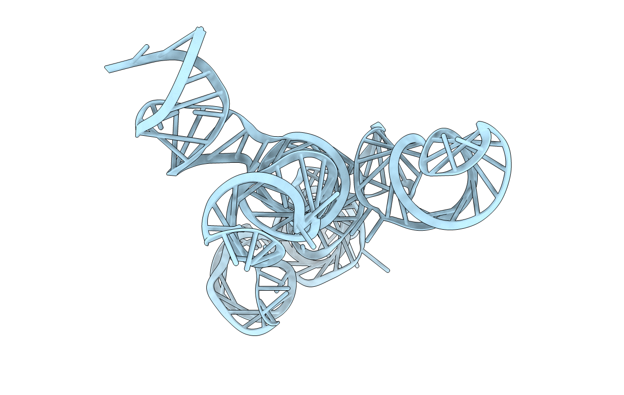
5V3I image
Entry Detail
PDB ID:
5V3I
Keywords:
Title:
Crystal structure of the VS ribozyme - wild-type C634
Biological Source:
Source Organism:
PDB Version:
Deposition Date:
2017-03-07
Release Date:
2017-07-05
Method Details:
Experimental Method:
Resolution:
3.29 Å
R-Value Free:
0.24
R-Value Work:
0.21
R-Value Observed:
0.21
Space Group:
P 43 21 2
Macromolecular Entities
Polymer Type:polyribonucleotide
Description:VS Ribozyme RNA
Chain IDs:A
Chain Length:186
Number of Molecules:1
Biological Source:Neurospora
Primary Citation
Structural Basis for Substrate Helix Remodeling and Cleavage Loop Activation in the Varkud Satellite Ribozyme.
J. Am. Chem. Soc. 139 9591 9597 (2017)
PMID: 28625058 DOI: 10.1021/jacs.7b03655

Abstact

The Varkud satellite (VS) ribozyme catalyzes site-specific RNA cleavage and ligation reactions. Recognition of the substrate involves a kissing loop interaction between the substrate and the catalytic domain of the ribozyme, resulting in a rearrangement of the substrate helix register into a so-called "shifted" conformation that is critical for substrate binding and activation. We report a 3.3 Å crystal structure of the complete ribozyme that reveals the active, shifted conformation of the substrate, docked into the catalytic domain of the ribozyme. Comparison to previous NMR structures of isolated, inactive substrates provides a physical description of substrate remodeling, and implicates roles for tertiary interactions in catalytic activation of the cleavage loop. Similarities to the hairpin ribozyme cleavage loop activation suggest general strategies to enhance fidelity in RNA folding and ribozyme cleavage.

Legend

Protein

Chemical

Disease

Primary Citation of related structures