5V1D image
Deposition Date 2017-03-02
Release Date 2018-02-14
Last Version Date 2024-03-20
Entry Detail
PDB ID:
5V1D
Title:
Complex structure of the bovine PERK luminal domain and its substrate peptide
Biological Source:
Source Organism:
Bos taurus (Taxon ID: 9913)
synthetic construct (Taxon ID: 32630)
Method Details:
Experimental Method:
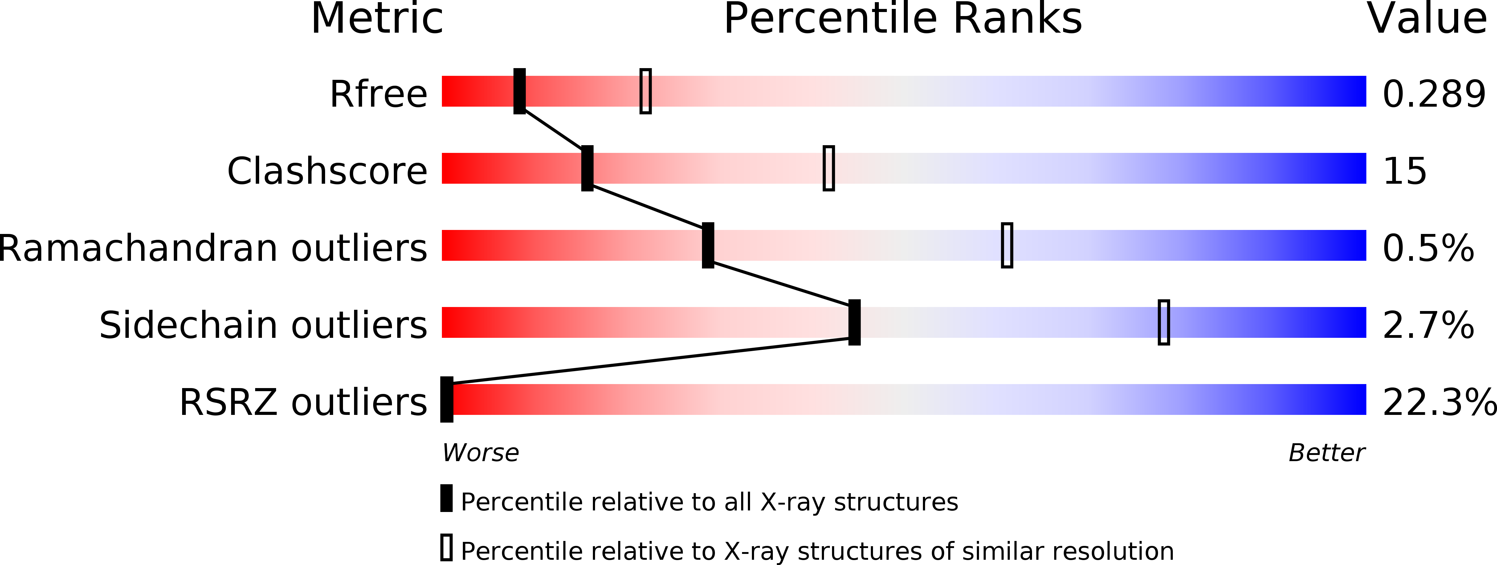
Resolution:
2.80 Å
R-Value Free:
0.29
R-Value Work:
0.24
R-Value Observed:
0.24
Space Group:
P 1 21 1
Macromolecular Entities
Structures with similar UniProt ID
Protein Blast
Polymer Type:polypeptide(L)
Molecule:eIF2AK3 protein
Gene (Uniprot):EIF2AK3
Mutagens:C337S
Chain IDs:A, C (auth: B), D (auth: C), F (auth: D)
Chain Length:326
Number of Molecules:4
Biological Source:Bos taurus
Polymer Type:polypeptide(L)
Molecule:12-mer peptide
Chain IDs:B (auth: E), E (auth: F), G
Chain Length:12
Number of Molecules:3
Biological Source:synthetic construct
Primary Citation
The luminal domain of the ER stress sensor protein PERK binds misfolded proteins and thereby triggers PERK oligomerization
J. Biol. Chem. 293 4110 4121 (2018)
PMID: 29386355 DOI: 10.1074/jbc.RA117.001294

Abstact

PRKR-like endoplasmic reticulum kinase (PERK) is one of the major sensor proteins that detect protein folding imbalances during endoplasmic reticulum (ER) stress. However, it remains unclear how ER stress activates PERK to initiate a downstream unfolded protein response (UPR). Here, we found that PERK's luminal domain can recognize and selectively interact with misfolded proteins but not with native proteins. Screening a phage-display library, we identified a peptide substrate, P16, of the PERK luminal domain and confirmed that P16 efficiently competes with misfolded proteins for binding this domain. To unravel the mechanism by which the PERK luminal domain interacts with misfolded proteins, we determined the crystal structure of the bovine PERK luminal domain complexed with P16 to 2.8-Å resolution. The structure revealed that PERK's luminal domain binds the peptide through a conserved hydrophobic groove. Substitutions within hydrophobic regions of the PERK luminal domain abolished the binding between PERK and misfolded proteins. We also noted that peptide binding results in major conformational changes in the PERK luminal domain that may favor PERK oligomerization. The structure of the PERK luminal domain-P16 complex suggested stacking of the luminal domain that leads to PERK oligomerization and activation via autophosphorylation after ligand binding. Collectively, our structural and biochemical results strongly support a ligand-driven model in which the PERK luminal domain interacts directly with misfolded proteins to induce PERK oligomerization and activation, resulting in ER stress signaling and the UPR.

Legend

Protein

Chemical

Disease

Primary Citation of related structures
Feedback Form
Name
Email
Institute
Feedback