5UX0 image
Deposition Date 2017-02-21
Release Date 2017-05-03
Last Version Date 2024-12-25
Entry Detail
PDB ID:
5UX0
Title:
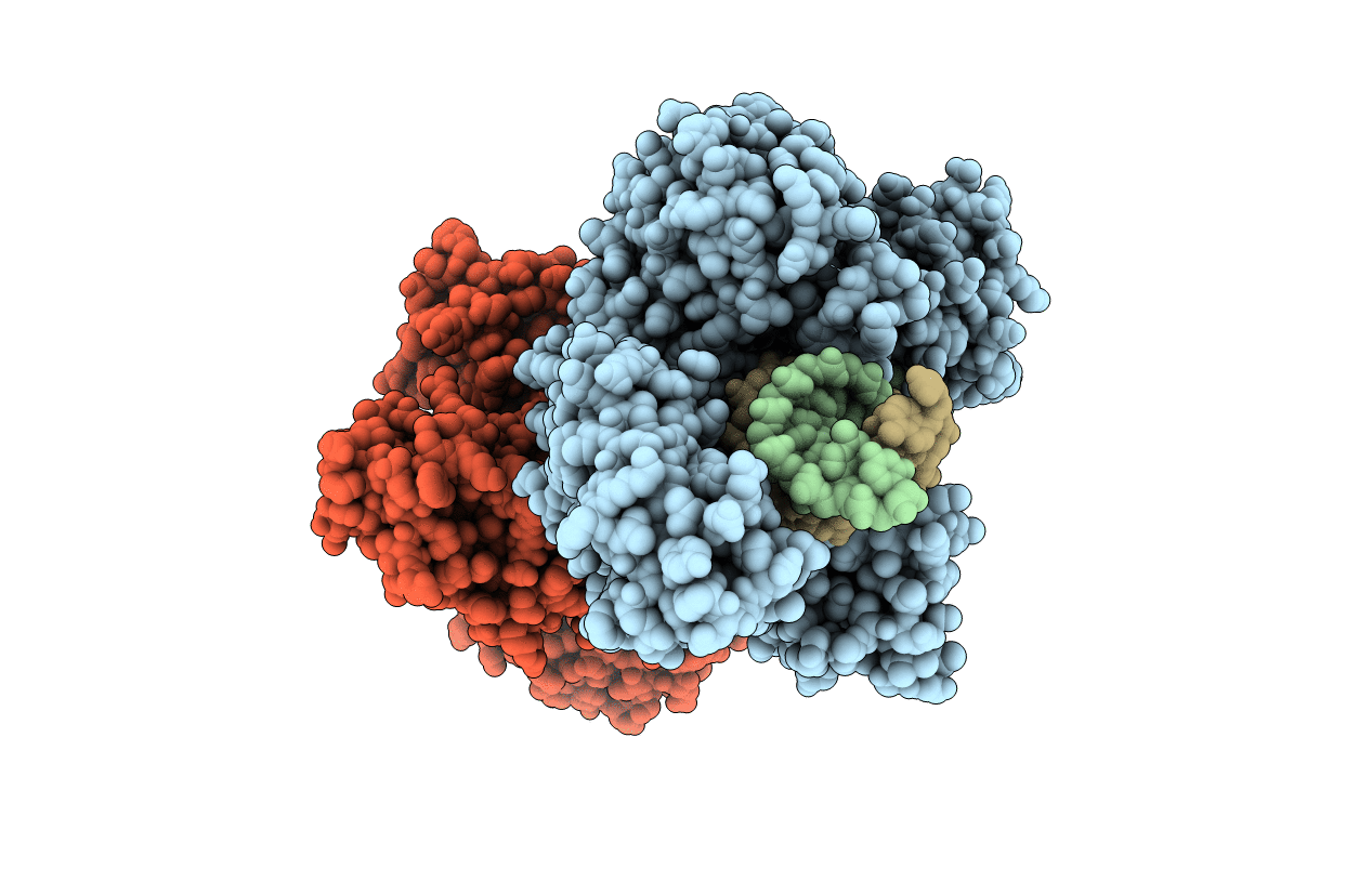
X-ray crystal structure of Marinitoga piezophila Argonaute in complex with 5' OH guide RNA and target DNA
Biological Source:
Source Organism:
Method Details:
Experimental Method:
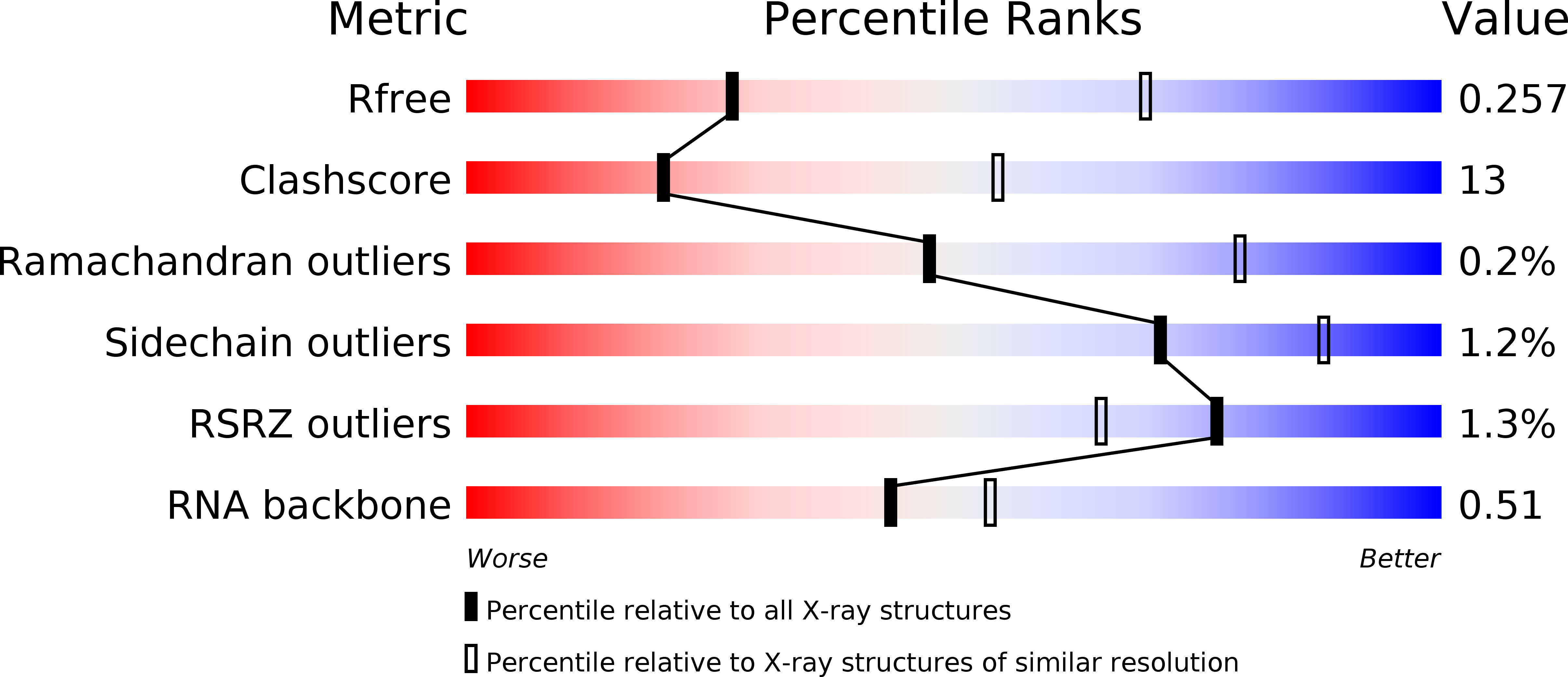
Resolution:
3.20 Å
R-Value Free:
0.25
R-Value Work:
0.21
R-Value Observed:
0.21
Space Group:
P 21 21 21
Macromolecular Entities
Structures with similar UniProt ID
Protein Blast
Polymer Type:polypeptide(L)
Molecule:Argonaute protein
Gene (Uniprot):ago
Mutagens:D516A
Chain IDs:A, D
Chain Length:642
Number of Molecules:2
Biological Source:Marinitoga piezophila
Polymer Type:polyribonucleotide
Molecule:RNA (5'-R(*G*GP*UP*AP*CP*AP*AP*CP*CP*UP*AP*CP*UP*AP*CP*CP*UP*CP*AP*U)-3')
Chain IDs:B, E
Chain Length:21
Number of Molecules:2
Biological Source:synthetic construct
Polymer Type:polydeoxyribonucleotide
Molecule:DNA (5'-D(P*AP*TP*GP*AP*GP*GP*TP*AP*GP*TP*AP*GP*GP*TP*TP*GP*TP*AP*CP*C)-3')
Chain IDs:C, F
Chain Length:21
Number of Molecules:2
Biological Source:synthetic construct
Ligand Molecules
Primary Citation
DNA recognition by an RNA-guided bacterial Argonaute.
PLoS ONE 12 e0177097 e0177097 (2017)
PMID: 28520746 DOI: 10.1371/journal.pone.0177097

Abstact

Argonaute (Ago) proteins are widespread in prokaryotes and eukaryotes and share a four-domain architecture capable of RNA- or DNA-guided nucleic acid recognition. Previous studies identified a prokaryotic Argonaute protein from the eubacterium Marinitoga piezophila (MpAgo), which binds preferentially to 5'-hydroxylated guide RNAs and cleaves single-stranded RNA (ssRNA) and DNA (ssDNA) targets. Here we present a 3.2 Å resolution crystal structure of MpAgo bound to a 21-nucleotide RNA guide and a complementary 21-nucleotide ssDNA substrate. Comparison of this ternary complex to other target-bound Argonaute structures reveals a unique orientation of the N-terminal domain, resulting in a straight helical axis of the entire RNA-DNA heteroduplex through the central cleft of the protein. Additionally, mismatches introduced into the heteroduplex reduce MpAgo cleavage efficiency with a symmetric profile centered around the middle of the helix. This pattern differs from the canonical mismatch tolerance of other Argonautes, which display decreased cleavage efficiency for substrates bearing sequence mismatches to the 5' region of the guide strand. This structural analysis of MpAgo bound to a hybrid helix advances our understanding of the diversity of target recognition mechanisms by Argonaute proteins.

Legend

Protein

Chemical

Disease

Primary Citation of related structures