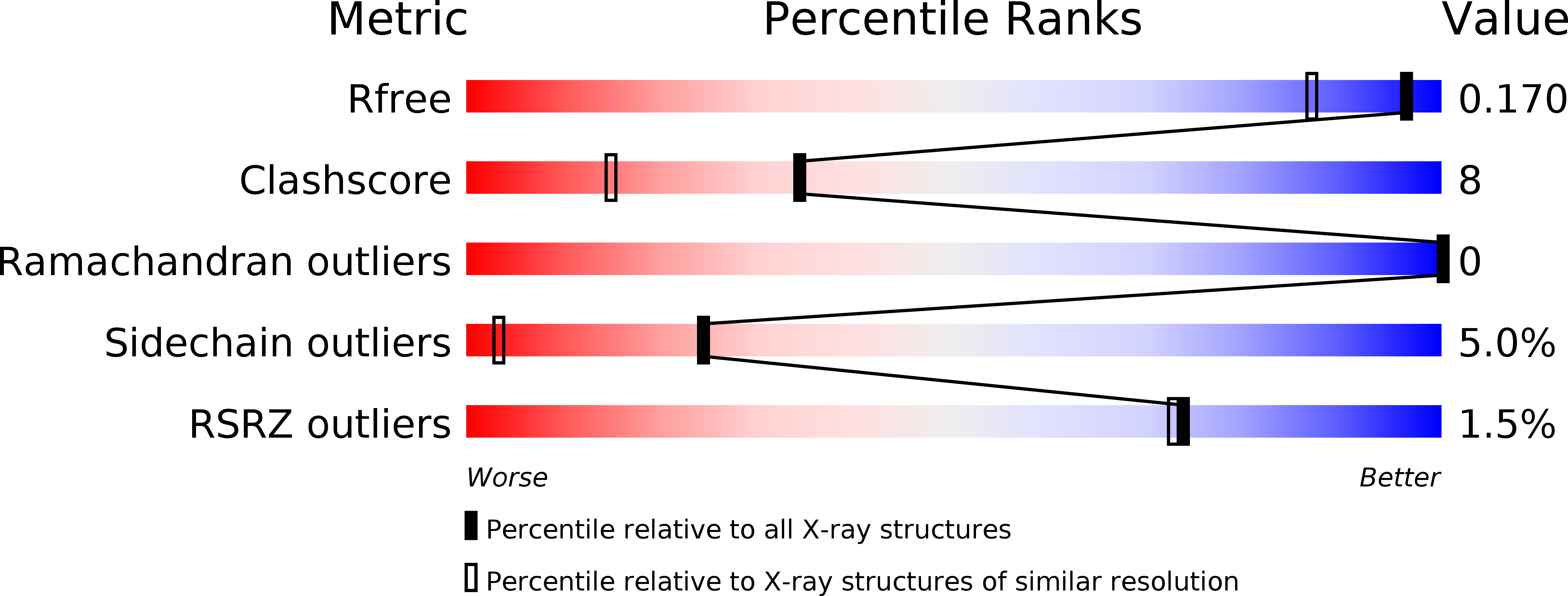
5UUI image
Deposition Date 2017-02-16
Release Date 2017-08-02
Last Version Date 2024-10-16
Entry Detail
PDB ID:
5UUI
Keywords:
Title:
Crystal Structure of Spin-Labeled T77C TNFa
Biological Source:
Source Organism:
Homo sapiens (Taxon ID: 9606)
Host Organism:
Method Details:
Experimental Method:
Resolution:
1.40 Å
R-Value Free:
0.17
R-Value Work:
0.13
R-Value Observed:
0.14
Space Group:
H 3
Macromolecular Entities
Structures with similar UniProt ID
Protein Blast
Polymer Type:polypeptide(L)
Molecule:Tumor necrosis factor
Gene (Uniprot):TNF
Chain IDs:A
Chain Length:158
Number of Molecules:1
Biological Source:Homo sapiens
Ligand Molecules
Primary Citation
Natural Conformational Sampling of Human TNF alpha Visualized by Double Electron-Electron Resonance.
Biophys. J. 113 371 380 (2017)
PMID: 28746848 DOI: 10.1016/j.bpj.2017.06.007

Abstact

Double electron-electron resonance in conjunction with site-directed spin labeling has been used to probe natural conformational sampling of the human tumor necrosis factor α trimer. We suggest a previously unreported, predeoligomerization conformation of the trimer that has been shown to be sampled at low frequency. A model of this trimeric state has been constructed based on crystal structures using the double-electron-electron-resonance distances. The model shows one of the protomers to be rotated and tilted outward at the tip end, leading to a breaking of the trimerous symmetry and distortion at a receptor-binding interface. The new structure offers opportunities to modulate the biological activity of tumor necrosis factor α through stabilization of the distorted trimer with small molecules.

Legend

Protein

Chemical

Disease

Primary Citation of related structures