Deposition Date
2017-02-08
Release Date
2017-05-10
Last Version Date
2023-10-04
Entry Detail
PDB ID:
5UQK
Keywords:
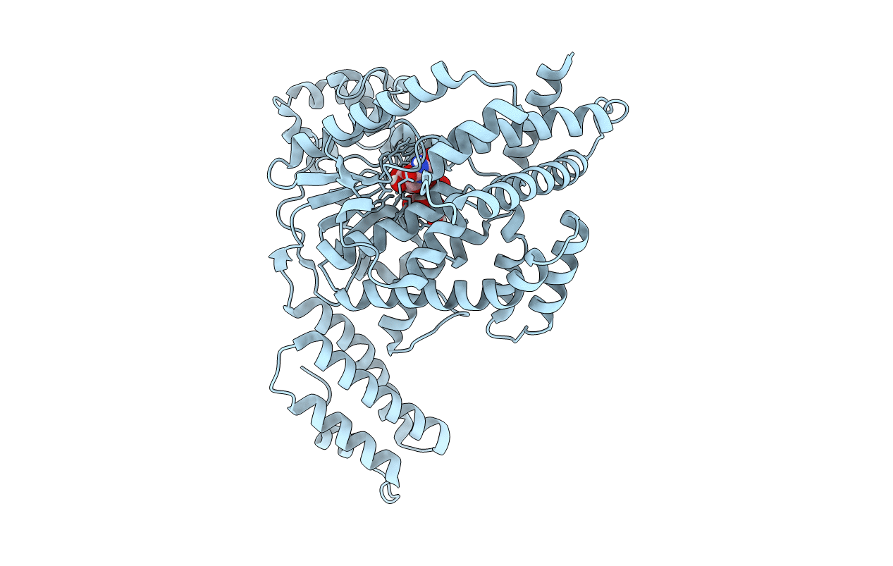
Title:
Clostridium difficile toxin A (TcdA) glucosyltransferase domain in complex with U2F
Biological Source:
Source Organism(s):
Clostridioides difficile (Taxon ID: 1496)
Expression System(s):
Method Details:
Experimental Method:
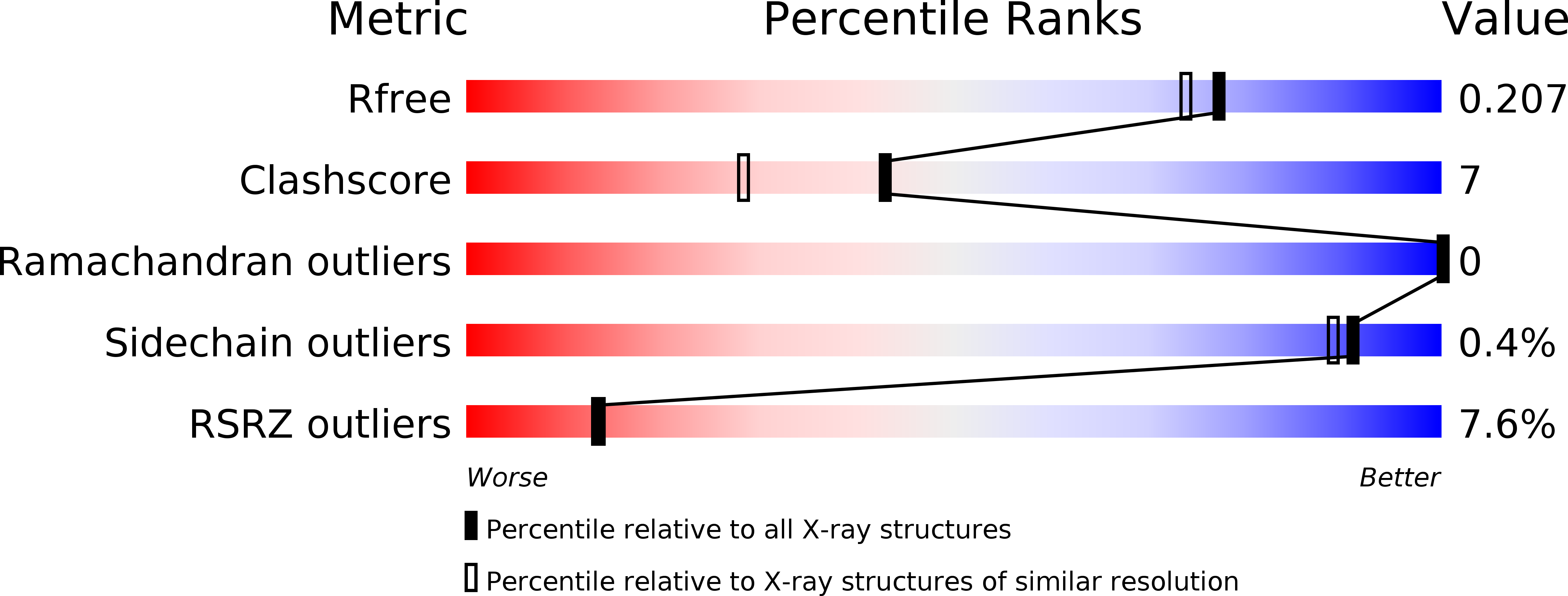
Resolution:
1.85 Å
R-Value Free:
0.20
R-Value Work:
0.18
R-Value Observed:
0.18
Space Group:
P 65