Deposition Date
2017-01-31
Release Date
2017-04-12
Last Version Date
2024-10-09
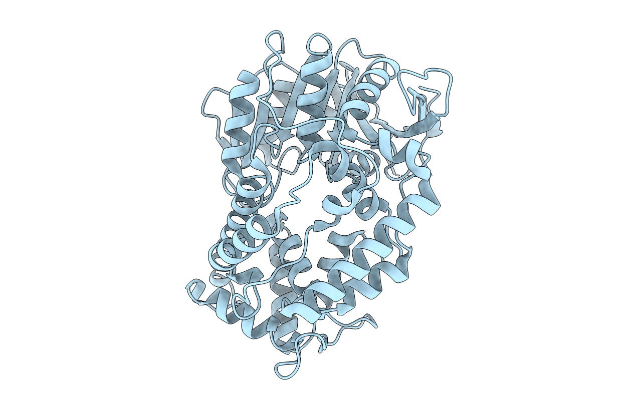
Entry Detail
Biological Source:
Source Organism(s):
Mycobacterium tuberculosis (Taxon ID: 83332)
Expression System(s):
Method Details:
Experimental Method:
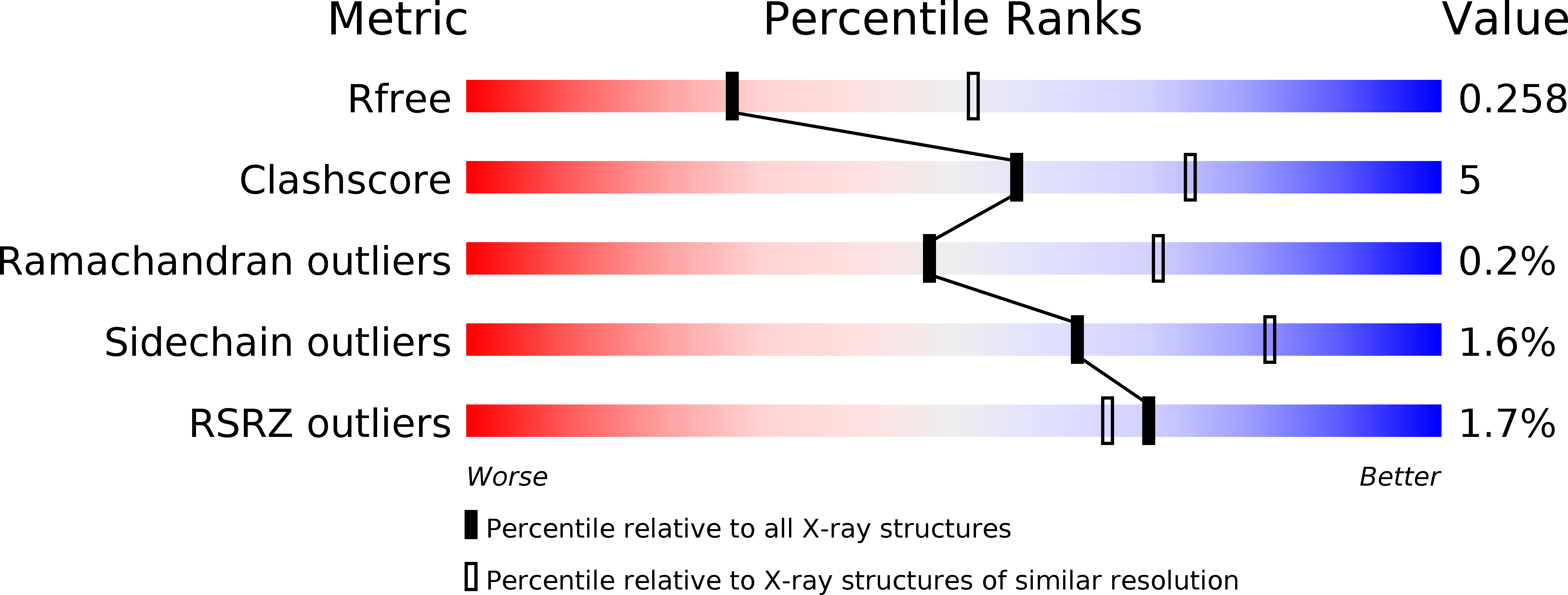
Resolution:
2.61 Å
R-Value Free:
0.25
R-Value Work:
0.21
R-Value Observed:
0.22
Space Group:
P 31 2 1