5UNJ image
Deposition Date 2017-01-31
Release Date 2017-04-19
Last Version Date 2023-10-04
Entry Detail
PDB ID:
5UNJ
Keywords:
Title:
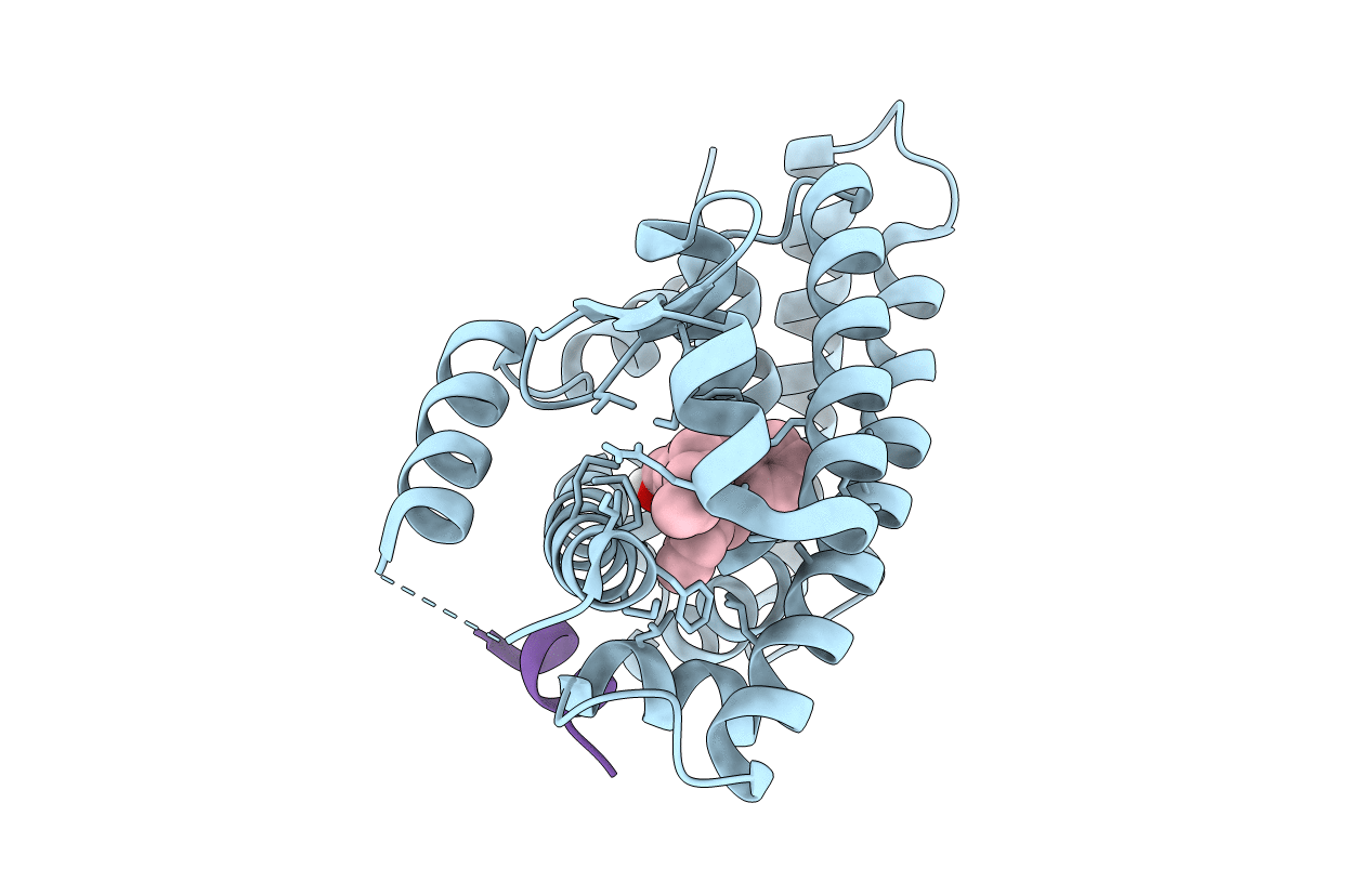
Structure of Human Liver Receptor Homolog 1 in complex with PGC1a and RJW100
Biological Source:
Source Organism:
Homo sapiens (Taxon ID: 9606)
Host Organism:
Method Details:
Experimental Method:
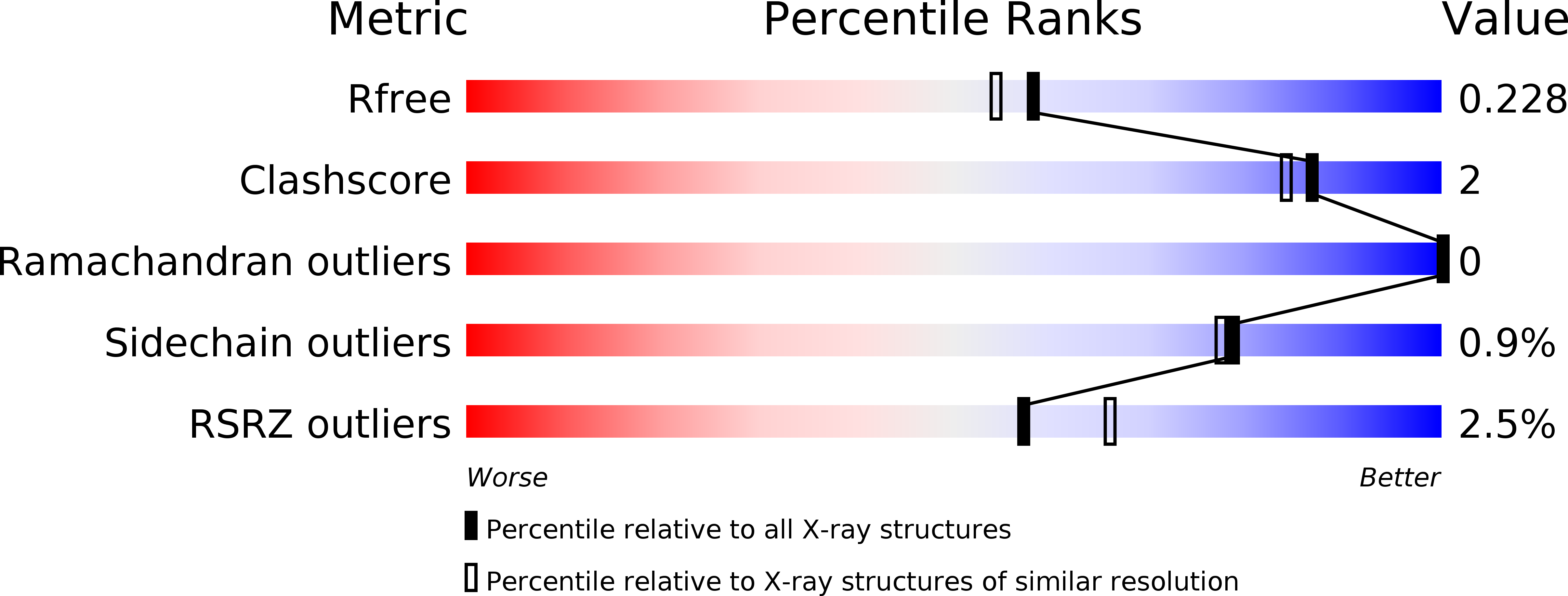
Resolution:
1.96 Å
R-Value Free:
0.22
R-Value Work:
0.19
R-Value Observed:
0.20
Space Group:
P 21 21 2
Macromolecular Entities
Structures with similar UniProt ID
Protein Blast
Polymer Type:polypeptide(L)
Molecule:Nuclear receptor subfamily 5 group A member 2
Gene (Uniprot):NR5A2
Chain IDs:A
Chain Length:245
Number of Molecules:1
Biological Source:Homo sapiens
Structures with similar UniProt ID
Protein Blast
Polymer Type:polypeptide(L)
Molecule:Peroxisome proliferator-activated gamma coactivator 1-alpha
Chain IDs:B (auth: C)
Chain Length:14
Number of Molecules:1
Biological Source:Homo sapiens
Ligand Molecules
Primary Citation
Structure and Dynamics of the Liver Receptor Homolog 1-PGC1 alpha Complex.
Mol. Pharmacol. 92 1 11 (2017)
PMID: 28363985 DOI: 10.1124/mol.117.108514

Abstact

Peroxisome proliferator-activated gamma coactivator 1-α (PGC1α) regulates energy metabolism by directly interacting with transcription factors to modulate gene expression. Among the PGC1α binding partners is liver receptor homolog 1 (LRH-1; NR5A2), an orphan nuclear hormone receptor that controls lipid and glucose homeostasis. Although PGC1α is known to bind and activate LRH-1, mechanisms through which PGC1α changes LRH-1 conformation to drive transcription are unknown. Here, we used biochemical and structural methods to interrogate the LRH-1-PGC1α complex. Purified, full-length LRH-1, as well as isolated ligand binding domain, bound to PGC1α with higher affinity than to the coactivator, nuclear receptor coactivator-2 (Tif2), in coregulator peptide recruitment assays. We present the first crystal structure of the LRH-1-PGC1α complex, which depicts several hydrophobic contacts and a strong charge clamp at the interface between these partners. In molecular dynamics simulations, PGC1α induced correlated atomic motion throughout the entire LRH-1 activation function surface, which was dependent on charge-clamp formation. In contrast, Tif2 induced weaker signaling at the activation function surface than PGC1α but promoted allosteric signaling from the helix 6/β-sheet region of LRH-1 to the activation function surface. These studies are the first to probe mechanisms underlying the LRH-1-PGC1α interaction and may illuminate strategies for selective therapeutic targeting of PGC1α-dependent LRH-1 signaling pathways.

Legend

Protein

Chemical

Disease

Primary Citation of related structures