5UN9 image
Entry Detail
PDB ID:
5UN9
Keywords:
Title:
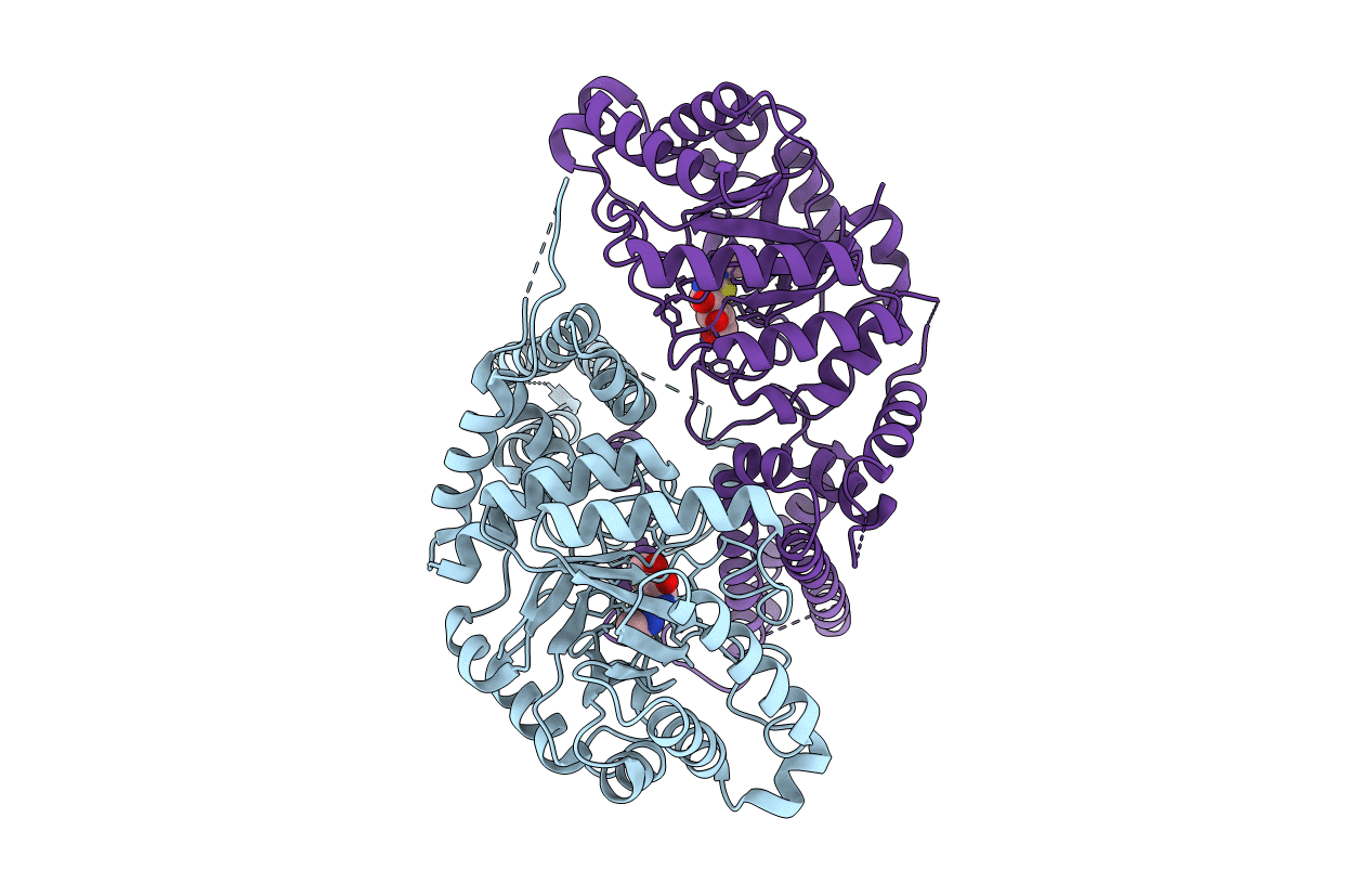
The crystal structure of human O-GlcNAcase in complex with Thiamet-G
Biological Source:
PDB Version:
Deposition Date:
2017-01-30
Release Date:
2017-03-15
Method Details:
Experimental Method:
Resolution:
2.50 Å
R-Value Free:
0.26
R-Value Work:
0.21
R-Value Observed:
0.21
Space Group:
P 1 21 1
Macromolecular Entities
Polymer Type:polypeptide(L)
Description:Protein O-GlcNAcase
Chain IDs:A, B
Chain Length:504
Number of Molecules:2
Biological Source:Homo sapiens
Ligand Molecules
Primary Citation
Structures of human O-GlcNAcase and its complexes reveal a new substrate recognition mode.
Nat. Struct. Mol. Biol. 24 362 369 (2017)
PMID: 28319083 DOI: 10.1038/nsmb.3390

Abstact

Human O-GlcNAcase (hOGA) is the unique enzyme responsible for the hydrolysis of the O-linked β-N-acetyl glucosamine (O-GlcNAc) modification, an essential protein glycosylation event that modulates the function of numerous cellular proteins in response to nutrients and stress. Here we report crystal structures of a truncated hOGA, which comprises the catalytic and stalk domains, in apo form, in complex with an inhibitor, and in complex with a glycopeptide substrate. We found that hOGA forms an unusual arm-in-arm homodimer in which the catalytic domain of one monomer is covered by the stalk domain of the sister monomer to create a substrate-binding cleft. Notably, the residues on the cleft surface afford extensive interactions with the peptide substrate in a recognition mode that is distinct from that of its bacterial homologs. These structures represent the first model of eukaryotic enzymes in the glycoside hydrolase 84 (GH84) family and provide a crucial starting point for understanding the substrate specificity of hOGA, which regulates a broad range of biological and pathological processes.

Legend

Protein

Chemical

Disease

Primary Citation of related structures