Deposition Date
2017-01-25
Release Date
2017-02-08
Last Version Date
2023-10-04
Entry Detail
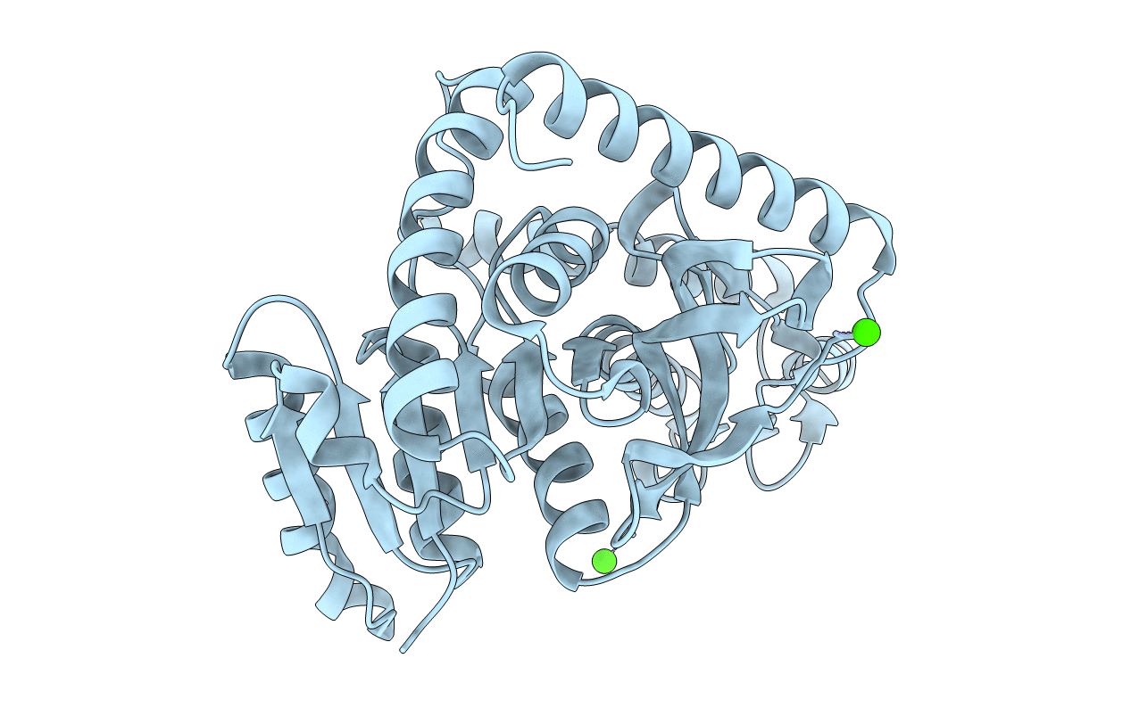
Biological Source:
Source Organism(s):
Methylobacterium extorquens (Taxon ID: 408)
Expression System(s):
Method Details:
Experimental Method:
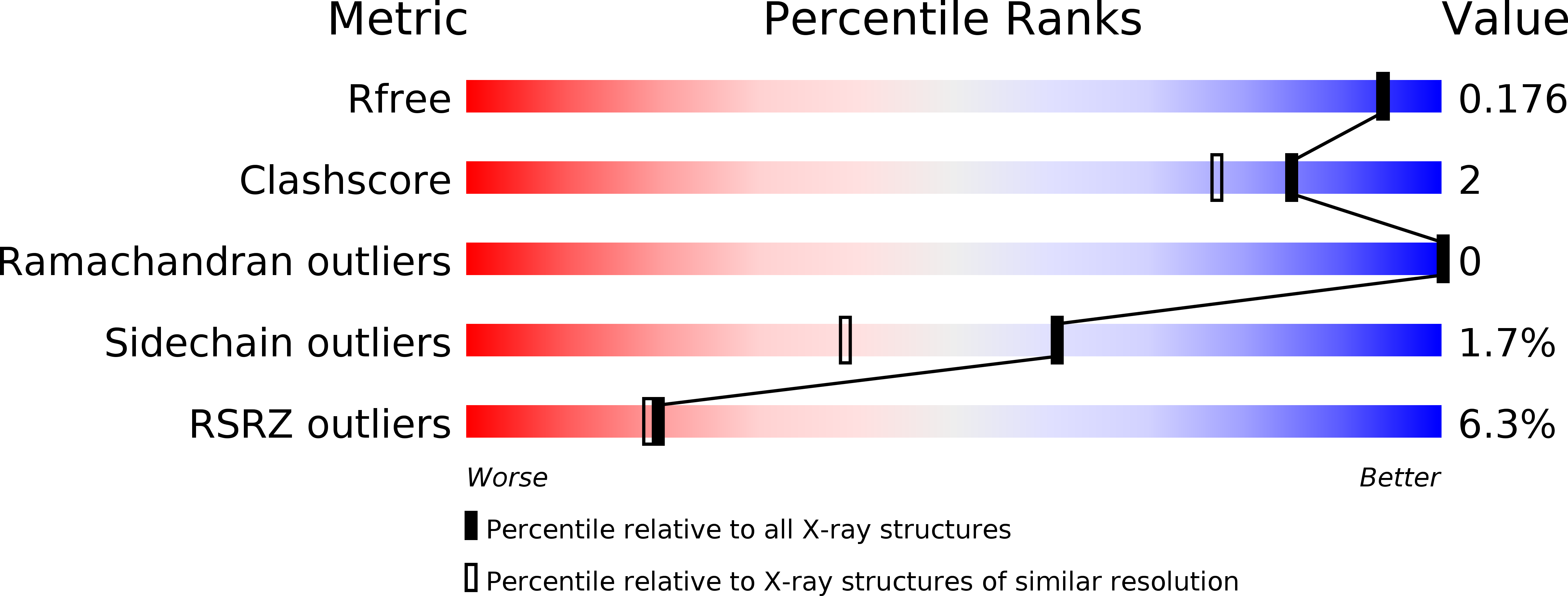
Resolution:
1.66 Å
R-Value Free:
0.16
R-Value Work:
0.13
R-Value Observed:
0.14
Space Group:
P 64 2 2