5UKW image
Deposition Date 2017-01-23
Release Date 2017-04-19
Last Version Date 2024-10-16
Entry Detail
PDB ID:
5UKW
Keywords:
Title:
Crystal structure of human Glucose 6-phosphate Dehydrogenase mutant (A277C) complexed with G6P
Biological Source:
Source Organism:
Homo sapiens (Taxon ID: 9606)
Host Organism:
Method Details:
Experimental Method:
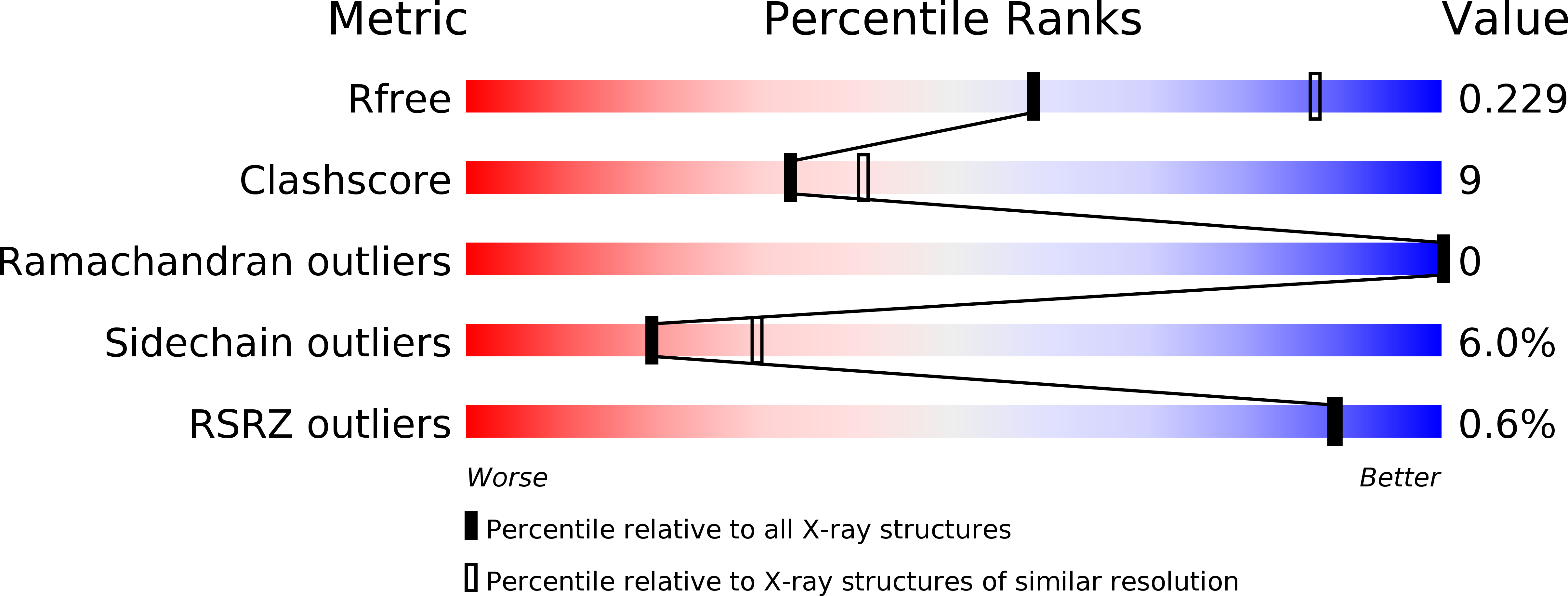
Resolution:
2.65 Å
R-Value Free:
0.23
R-Value Work:
0.18
R-Value Observed:
0.19
Space Group:
F 2 2 2
Macromolecular Entities
Structures with similar UniProt ID
Protein Blast
Polymer Type:polypeptide(L)
Molecule:Glucose-6-phosphate 1-dehydrogenase
Gene (Uniprot):G6PD
Mutagens:Q28F, A277C
Chain IDs:A
Chain Length:488
Number of Molecules:1
Biological Source:Homo sapiens
Primary Citation
Mutations in the tetramer interface of human glucose-6-phosphate dehydrogenase reveals kinetic differences between oligomeric states.
FEBS Lett. 591 1278 1284 (2017)
PMID: 28370139 DOI: 10.1002/1873-3468.12638

Abstact

Glucose-6-phosphate dehydrogenase (G6PDH) catalyzes the oxidation of glucose-6-phoshate to 6-phospho-gluconolactone with the concomitant reduction of NADP+ to NADPH. In solution, the recombinant human G6PDH is known to be active as dimers and tetramers. To distinguish between the kinetic properties of dimers and tetramers of the G6PDH is not trivial. Steady-state kinetic experiments are often performed at low enzyme concentrations, which favor the dimeric state. The present work describes two novel human G6PDH mutants, one that creates four disulfide bonds among apposing dimers, resulting in a 'cross-linked' tetramer, and another that prevents the dimer to dimer association. The functional and structural characterizations of such mutants indicate the tetramer as the most active form of human G6PDH.

Legend

Protein

Chemical

Disease

Primary Citation of related structures
Feedback Form
Name
Email
Institute
Feedback