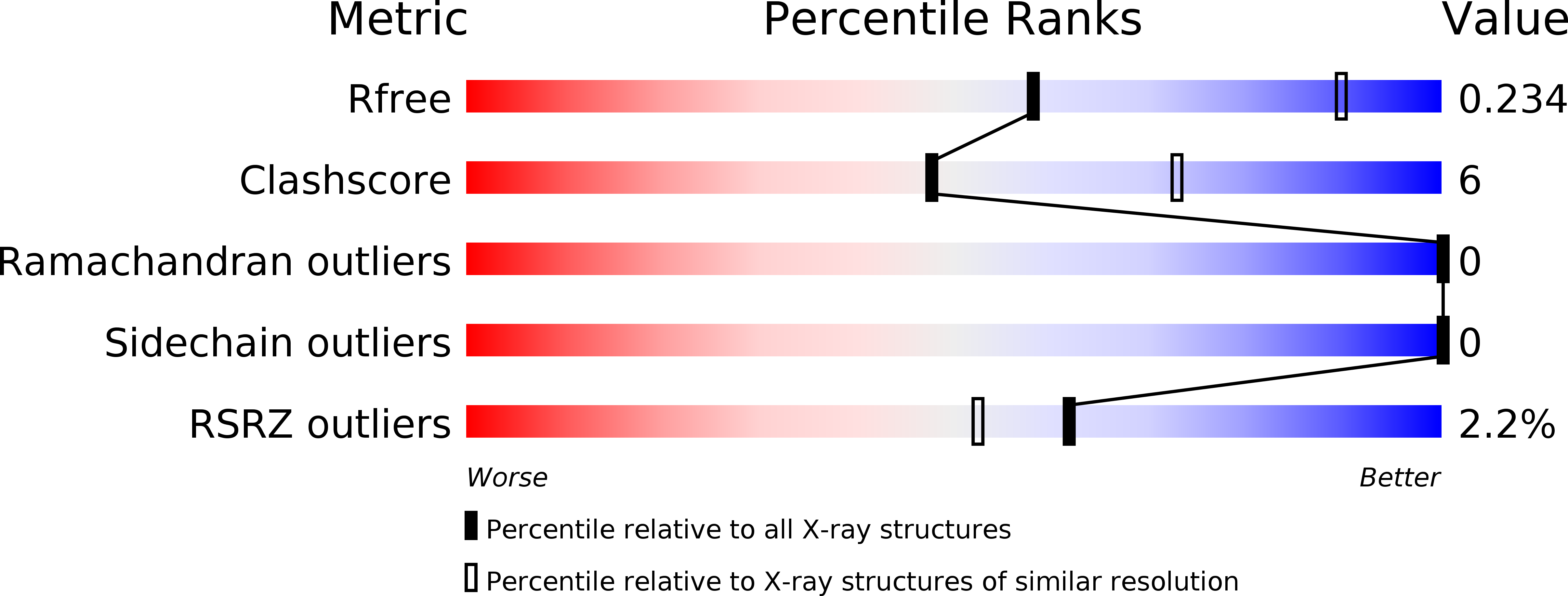
5UK3 image
Deposition Date 2017-01-19
Release Date 2017-06-21
Last Version Date 2023-10-04
Entry Detail
PDB ID:
5UK3
Keywords:
Title:
Crystal structure of cyanase from T. urticae
Biological Source:
Source Organism:
Host Organism:
Method Details:
Experimental Method:
Resolution:
2.80 Å
R-Value Free:
0.23
R-Value Work:
0.22
R-Value Observed:
0.22
Space Group:
P 1 21 1
Macromolecular Entities
Polymer Type:polypeptide(L)
Molecule:Uncharacterized protein
Gene (Uniprot):107368746
Chain IDs:A, B, C, D, E, F, G, H, I, J
Chain Length:176
Number of Molecules:10
Biological Source:Tetranychus urticae
Primary Citation
Structural Characterization of a Eukaryotic Cyanase from Tetranychus urticae.
J. Agric. Food Chem. 65 5453 5462 (2017)
PMID: 28613863 DOI: 10.1021/acs.jafc.7b01333

Abstact

The two-spotted spider mite Tetranychus urticae is a polyphagous agricultural pest and poses a high risk to global crop production as it is rapidly developing pesticide resistance. Genomic and transcriptomic analysis has revealed the presence of a remarkable cyanase gene in T. urticae and related mite species within the Acariformes lineage. Cyanase catalyzes the detoxification of cyanate and is potentially an attractive protein target for the development of new acaricides. Phylogenetic analysis indicates that within the Acariformes, the cyanase gene originates from a single horizontal gene transfer event, which precedes subsequent speciation. Our structural studies presented here compare and contrast prokaryotic cyanases to T. urticae cyanase, which all form homodecamers and have conserved active site residues, but display different surface areas between homodimers in the overall decameric structure.

Legend

Protein

Chemical

Disease

Primary Citation of related structures