5UJ5 image
Entry Detail
PDB ID:
5UJ5
Title:
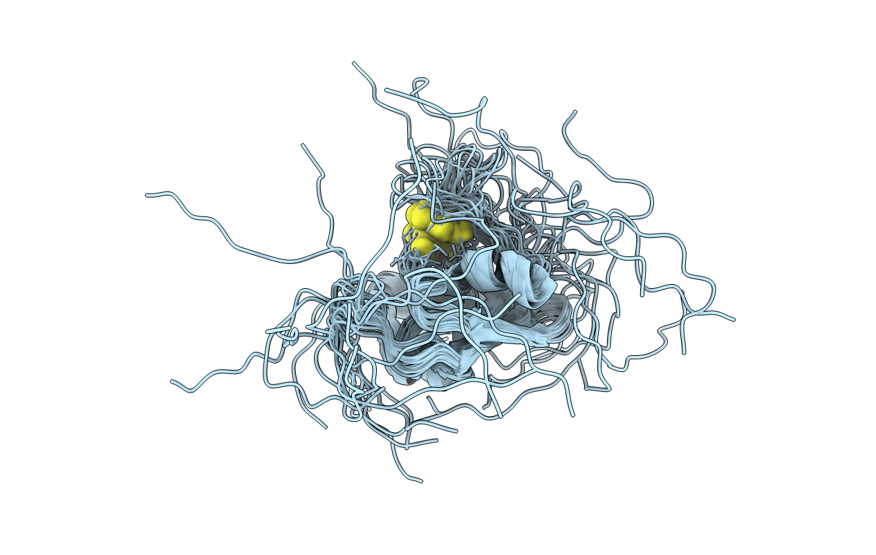
Solution structure of the oxidized iron-sulfur protein adrenodoxin from Encephalitozoon cuniculi. Seattle Structural Genomics Center for Infectious Disease target EncuA.00705.a
Biological Source:
Source Organism:
Host Organism:
PDB Version:
Deposition Date:
2017-01-17
Release Date:
2017-02-01
Method Details:
Experimental Method:
Conformers Calculated:
100
Conformers Submitted:
20
Selection Criteria:
target function
Macromolecular Entities
Polymer Type:polypeptide(L)
Description:Adrenodoxin
Chain IDs:A
Chain Length:132
Number of Molecules:1
Biological Source:Encephalitozoon cuniculi
Ligand Molecules
Primary Citation
Solution structure for an Encephalitozoon cuniculi adrenodoxin-like protein in the oxidized state.
Protein Sci. 29 809 817 (2020)
PMID: 31912584 DOI: 10.1002/pro.3818

Abstact

Encephalitozoon cuniculi is a unicellular, obligate intracellular eukaryotic parasite in the Microsporidia family and one of the agents responsible for microsporidosis infections in humans. Like most Microsporidia, the genome of E. cuniculi is markedly reduced and the organism contains mitochondria-like organelles called mitosomes instead of mitochondria. Here we report the solution NMR structure for a protein physically associated with mitosome-like organelles in E. cuniculi, the 128-residue, adrenodoxin-like protein Ec-Adx (UniProt ID Q8SV19) in the [2Fe-2S] ferredoxin superfamily. Oxidized Ec-Adx contains a mixed four-strand β-sheet, β2-β1-β4-β3 (↓↑↑↓), loosely encircled by three α-helices and two 310 -helices. This fold is similar to the structure observed in other adrenodoxin and adrenodoxin-like proteins except for the absence of a fifth anti-parallel β-strand next to β3 and the position of α3. Cross peaks are missing or cannot be unambiguously assigned for 20 amide resonances in the 1 H-15 N HSQC spectrum of Ec-Adx. These missing residues are clustered primarily in two regions, G48-V61 and L94-L98, containing the four cysteine residues predicted to ligate the paramagnetic [2Fe-2S] cluster. Missing amide resonances in 1 H-15 N HSQC spectra are detrimental to NMR-based solution structure calculations because 1 H-1 H NOE restraints are absent (glass half-empty) and this may account for the absent β-strand (β5) and the position of α3 in oxidized Ec-Adx. On the other hand, the missing amide resonances unambiguously identify the presence, and immediate environment, of the paramagnetic [2Fe-2S] cluster in oxidized Ec-Adx (glass half-full).

Legend

Protein

Chemical

Disease

Primary Citation of related structures